Copyright
©2012 Baishideng Publishing Group Co.
World J Radiol. Jun 28, 2012; 4(6): 258-264
Published online Jun 28, 2012. doi: 10.4329/wjr.v4.i6.258
Published online Jun 28, 2012. doi: 10.4329/wjr.v4.i6.258
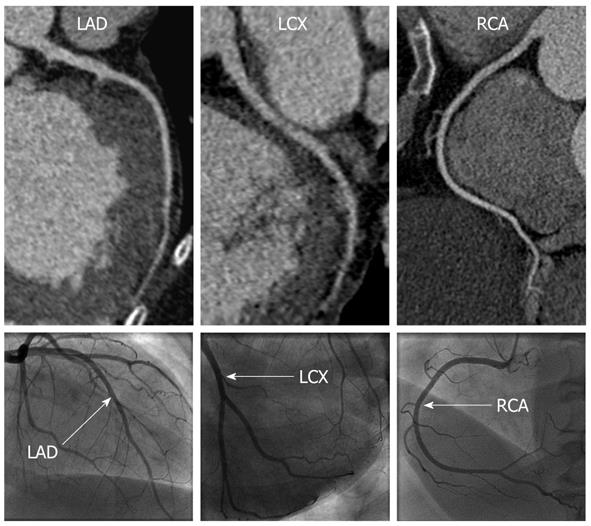
Figure 2 A 58-year-old woman with dilated cardiomyopathy with no significant coronary artery disease.
Curved reformations of the coronary computed tomography angiography images (top) and corresponding X-ray coronary angiography images (bottom) are shown for the left anterior descending (LAD), left circumflex (LCX) and right coronary artery (RCA).
- Citation: Srichai MB, Fisch M, Hecht E, Slater J, Rachofsky E, Hays AG, Babb J, Jacobs JE. Dual source computed tomography coronary angiography in new onset cardiomyopathy. World J Radiol 2012; 4(6): 258-264
- URL: https://www.wjgnet.com/1949-8470/full/v4/i6/258.htm
- DOI: https://dx.doi.org/10.4329/wjr.v4.i6.258


