Copyright
©2012 Baishideng Publishing Group Co.
World J Radiol. Jun 28, 2012; 4(6): 258-264
Published online Jun 28, 2012. doi: 10.4329/wjr.v4.i6.258
Published online Jun 28, 2012. doi: 10.4329/wjr.v4.i6.258
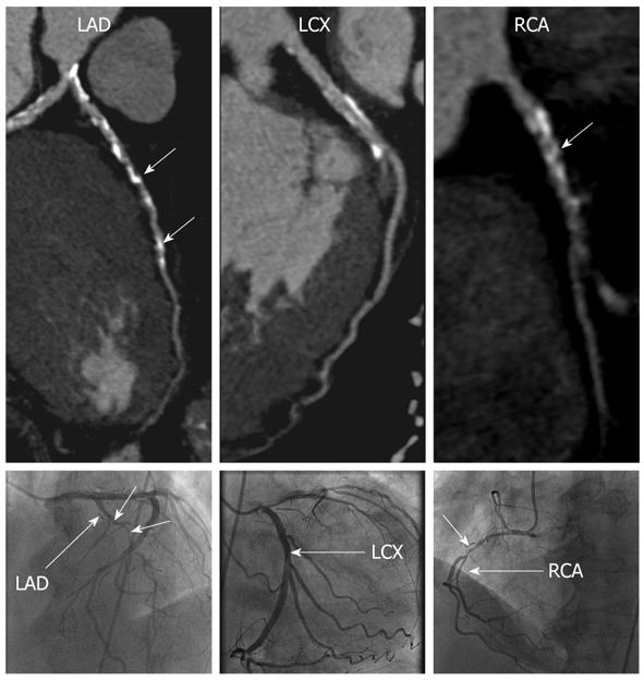
Figure 1 A 78-year-old man with dilated cardiomyopathy associated with severe coronary artery disease.
Curved reformations of the coronary computed tomography angiography images (top) and corresponding X-ray coronary angiography images (bottom) are shown for the left anterior descending (LAD), left circumflex (LCX) and right coronary artery (RCA). Focal areas of severe stenoses (short arrows) are noted in the LAD and RCA.
- Citation: Srichai MB, Fisch M, Hecht E, Slater J, Rachofsky E, Hays AG, Babb J, Jacobs JE. Dual source computed tomography coronary angiography in new onset cardiomyopathy. World J Radiol 2012; 4(6): 258-264
- URL: https://www.wjgnet.com/1949-8470/full/v4/i6/258.htm
- DOI: https://dx.doi.org/10.4329/wjr.v4.i6.258


