Copyright
©2012 Baishideng Publishing Group Co.
World J Radiol. May 28, 2012; 4(5): 231-235
Published online May 28, 2012. doi: 10.4329/wjr.v4.i5.231
Published online May 28, 2012. doi: 10.4329/wjr.v4.i5.231
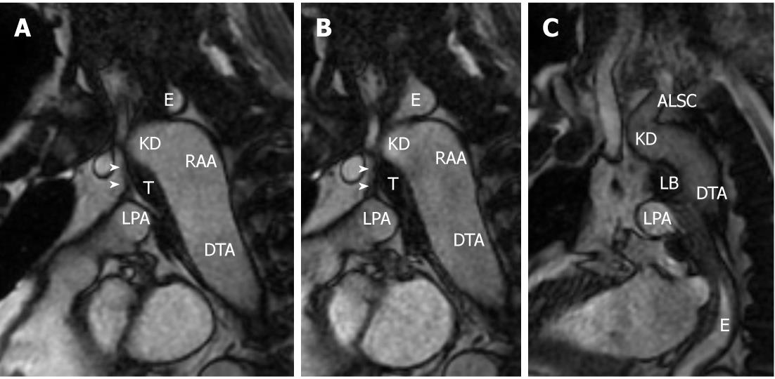
Figure 4 Three sequential frames of a cine-FIESTA sagittal oblique sequence.
A, B: Progressive distension of the esophageal lumen superiorly to the right aortic arch (RAA); C: The water bolus is appreciable in the lower thoracic esophagus, but it was impossible to obtain a significant distention of the esophageal segment passing through the vascular ring formed in its left lateral inferior part by the ligamentum arteriosum. Arrowheads indicate ligamentum arteriosum. T: Trachea; E: Esophagus; ALSC: Aberrant left subclavian artery; KD: Kommerell’s diverticulum; DTA: Descending thoracic aorta; LPA: Left pulmonary artery.
- Citation: Paparo F, Bacigalupo L, Melani E, Rollandi GA, De Caro G. Cardiac-MRI demonstration of the ligamentum arteriosum in a case of right aortic arch with aberrant left subclavian artery. World J Radiol 2012; 4(5): 231-235
- URL: https://www.wjgnet.com/1949-8470/full/v4/i5/231.htm
- DOI: https://dx.doi.org/10.4329/wjr.v4.i5.231


