Copyright
©2012 Baishideng Publishing Group Co.
World J Radiol. May 28, 2012; 4(5): 231-235
Published online May 28, 2012. doi: 10.4329/wjr.v4.i5.231
Published online May 28, 2012. doi: 10.4329/wjr.v4.i5.231
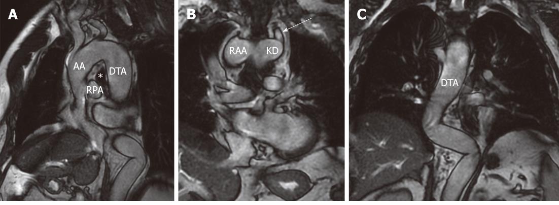
Figure 2 Cardiac-gated FIESTA images.
A: Sagittal oblique cardiac-gated FIESTA image (A) demonstrating the right aortic arch above the right main bronchus (asterisk) and right main pulmonary artery; B, C: Coronal cardiac-gated FIESTA images showing the relationships between the RAA and Kommerell's diverticulum from which the ALSC (large arrow in B) originates. The descending thoracic aorta courses on the right side of the spine and eventually turns on the left side to enter the aortic diaphragmatic hiatus. Large arrow indicate aberrant left subclavian artery. AA: Ascending aorta; DTA: Descending thoracic aorta; *: Right main bronchus; RPA: Right pulmonary artery; RAA: Right aortic arch; KD: Kommerell’s diverticulum.
- Citation: Paparo F, Bacigalupo L, Melani E, Rollandi GA, De Caro G. Cardiac-MRI demonstration of the ligamentum arteriosum in a case of right aortic arch with aberrant left subclavian artery. World J Radiol 2012; 4(5): 231-235
- URL: https://www.wjgnet.com/1949-8470/full/v4/i5/231.htm
- DOI: https://dx.doi.org/10.4329/wjr.v4.i5.231


