Copyright
©2012 Baishideng Publishing Group Co.
World J Radiol. May 28, 2012; 4(5): 224-227
Published online May 28, 2012. doi: 10.4329/wjr.v4.i5.224
Published online May 28, 2012. doi: 10.4329/wjr.v4.i5.224
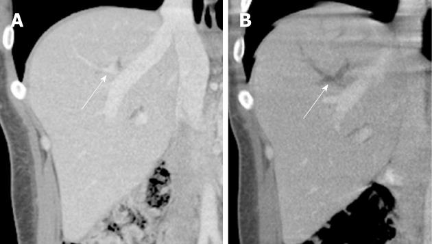
Figure 2 Contrast-enhanced computed tomography images before the percutaneous transhepatic cholangiography demonstrate a patent Segment VIII portal vein branch that developed localized portal vein thrombosis after the procedure.
A: A coronal reformatted image from a contrast-enhanced computed tomography (CT) image demonstrates that the portal vein branches (white arrow) to Segment VIII are patent; B: A coronal reformatted images from the contrast-enhanced CT scan performed when the patient presented to the emergency room 48 h after the percutaneous transhepatic cholangiography (PTC) procedure demonstrates thrombosis of a segmental and several subsegmental portal vein branches (white arrow) in Segment VIII, in the same location where the PTC was performed.
- Citation: Brennan IM, Ahmed M. Portal vein thrombosis following percutaneous transhepatic cholangiography-An unusual presentation of Prothrombin (Factor II) gene mutation. World J Radiol 2012; 4(5): 224-227
- URL: https://www.wjgnet.com/1949-8470/full/v4/i5/224.htm
- DOI: https://dx.doi.org/10.4329/wjr.v4.i5.224


