Copyright
©2012 Baishideng Publishing Group Co.
World J Radiol. May 28, 2012; 4(5): 224-227
Published online May 28, 2012. doi: 10.4329/wjr.v4.i5.224
Published online May 28, 2012. doi: 10.4329/wjr.v4.i5.224
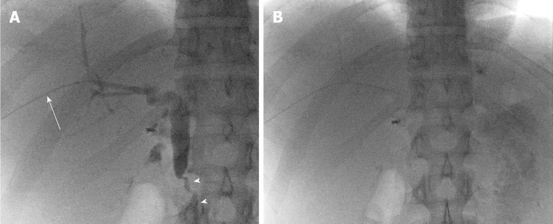
Figure 1 Fluoroscopic spot image from the second percutaneous transhepatic cholangiography procedure performed in the right lobe.
A: On this fluoroscopic spot image from the second percutaneous transhepatic cholangiography procedure performed through the right biliary system, the small 3Fr inner cannula (white arrow) is seen traversing the right hepatic lobe. Contrast opacification demonstrates filling of the right intrahepatic ducts and common bile duct, all normal in caliber. There is also brisk flow through the ampulla into the duodenum (white arrowheads); B: Delayed fluoroscopic image demonstrates complete emptying of the biliary system with contrast flowing forward into the duodenum and jejunum, without evidence of a biliary stricture. The 3Fr inner cannula was then removed and tract embolization was performed without incident.
- Citation: Brennan IM, Ahmed M. Portal vein thrombosis following percutaneous transhepatic cholangiography-An unusual presentation of Prothrombin (Factor II) gene mutation. World J Radiol 2012; 4(5): 224-227
- URL: https://www.wjgnet.com/1949-8470/full/v4/i5/224.htm
- DOI: https://dx.doi.org/10.4329/wjr.v4.i5.224


