Copyright
©2012 Baishideng Publishing Group Co.
World J Radiol. May 28, 2012; 4(5): 186-192
Published online May 28, 2012. doi: 10.4329/wjr.v4.i5.186
Published online May 28, 2012. doi: 10.4329/wjr.v4.i5.186
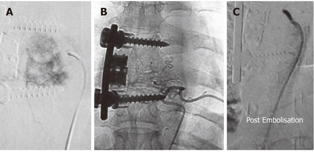
Figure 5 Pre-operative trans-arterial Embolisation of a thoracic vertebral hemangioma in a young male.
A: Twenty-three-year-old male with pain in the upper back for 3 mo. Radiographs revealed an expansile lytic lesion of the left transverse process of the sixth thoracic vertebra. Computed tomographic-guided biopsy of the lesion confirmed it to be a hemangioma. A flush aortogram with an angiographic pigtail placed at the level of the aortic arch identified the tumor feeding artery arising from the sixth left posterior intercostal artery which was selectively cannulated; B: Selective angiogram obtained from the feeder revealed marked tumor blush; C: Angiogram obtained after Embolisation with gelfoam showed almost complete loss of tumor blush.
- Citation: Gupta P, Gamanagatti S. Preoperative transarterial Embolisation in bone tumors. World J Radiol 2012; 4(5): 186-192
- URL: https://www.wjgnet.com/1949-8470/full/v4/i5/186.htm
- DOI: https://dx.doi.org/10.4329/wjr.v4.i5.186


