Copyright
©2012 Baishideng Publishing Group Co.
World J Radiol. Apr 28, 2012; 4(4): 179-185
Published online Apr 28, 2012. doi: 10.4329/wjr.v4.i4.179
Published online Apr 28, 2012. doi: 10.4329/wjr.v4.i4.179
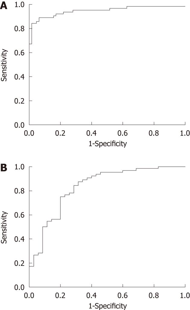
Figure 2 Receiver operating characteristic curve of difference in contrast enhancement between esophageal squamous cell carcinoma and background normal esophagus.
A: Discriminating the tumor from background normal esophagus (area under ROC curve = 0.948, P < 0.0001); B: Discriminating between the therapeutic change of esophageal squamous carcinoma treated with and without chemoradiotherapy (area under ROC curve = 0.833, P < 0.0001).
- Citation: Li R, Chen TW, Wang LY, Zhou L, Li H, Chen XL, Li CP, Zhang XM, Xiao RH. Quantitative measurement of contrast enhancement of esophageal squamous cell carcinoma on clinical MDCT. World J Radiol 2012; 4(4): 179-185
- URL: https://www.wjgnet.com/1949-8470/full/v4/i4/179.htm
- DOI: https://dx.doi.org/10.4329/wjr.v4.i4.179


