Copyright
©2012 Baishideng Publishing Group Co.
World J Radiol. Apr 28, 2012; 4(4): 167-173
Published online Apr 28, 2012. doi: 10.4329/wjr.v4.i4.167
Published online Apr 28, 2012. doi: 10.4329/wjr.v4.i4.167
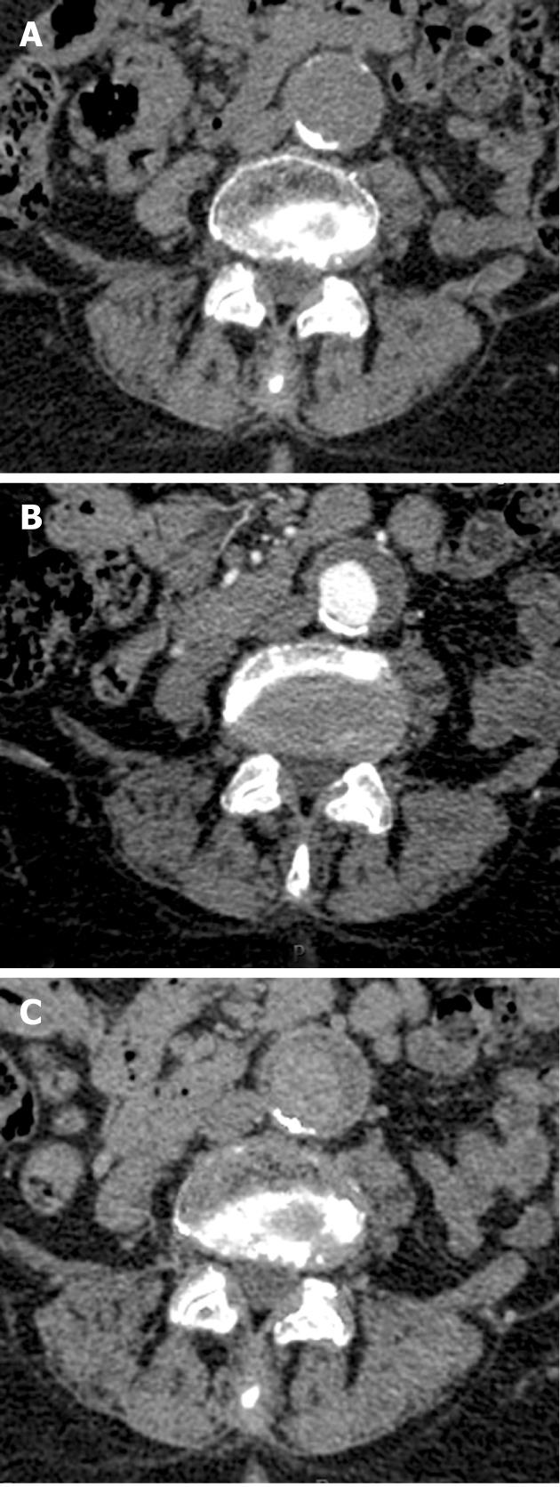
Figure 2 A 60-year-old male with an abdominal aortic aneurysm.
A: Regular nonenhanced image demonstrates a curved calcification in the posterior border of the aneurysm with some smaller calcifications in the anterior border; B: The contrast enhanced image shows opacification of the lumen; C: Virtual nonenhanced shows preserved posterior calcifications with deletion of the smaller anterior calcifications.
- Citation: Sosna J, Mahgerefteh S, Goshen L, Kafri G, Aviram G, Blachar A. Virtual nonenhanced abdominal dual-energy MDCT: Analysis of image characteristics. World J Radiol 2012; 4(4): 167-173
- URL: https://www.wjgnet.com/1949-8470/full/v4/i4/167.htm
- DOI: https://dx.doi.org/10.4329/wjr.v4.i4.167


