Copyright
©2012 Baishideng Publishing Group Co.
World J Radiol. Apr 28, 2012; 4(4): 151-158
Published online Apr 28, 2012. doi: 10.4329/wjr.v4.i4.151
Published online Apr 28, 2012. doi: 10.4329/wjr.v4.i4.151
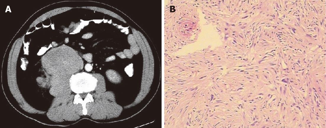
Figure 7 A 59-year-old male with retroperitoneal psoas muscle malignant fibrous histiocytoma.
A: Contrast-enhanced computed tomography scan arterial phase shows mass in retroperitoneum with clear margin, no obvious enhancement of mass and area of low density corresponding to necrosis. Note that the fat plane between the mass and psoas muscle is not evident, and the mass seems to be in direct contact with the right psoas muscle; B: Pathological specimen HE stain (× 400) shows spindle to oval tumor cells arranged in a criss-cross fashion.
- Citation: Karki B, Xu YK, Wu YK, Zhang WW. Primary malignant fibrous histiocytoma of the abdominal cavity: CT findings and pathological correlation. World J Radiol 2012; 4(4): 151-158
- URL: https://www.wjgnet.com/1949-8470/full/v4/i4/151.htm
- DOI: https://dx.doi.org/10.4329/wjr.v4.i4.151


