Copyright
©2012 Baishideng Publishing Group Co.
World J Radiol. Apr 28, 2012; 4(4): 151-158
Published online Apr 28, 2012. doi: 10.4329/wjr.v4.i4.151
Published online Apr 28, 2012. doi: 10.4329/wjr.v4.i4.151
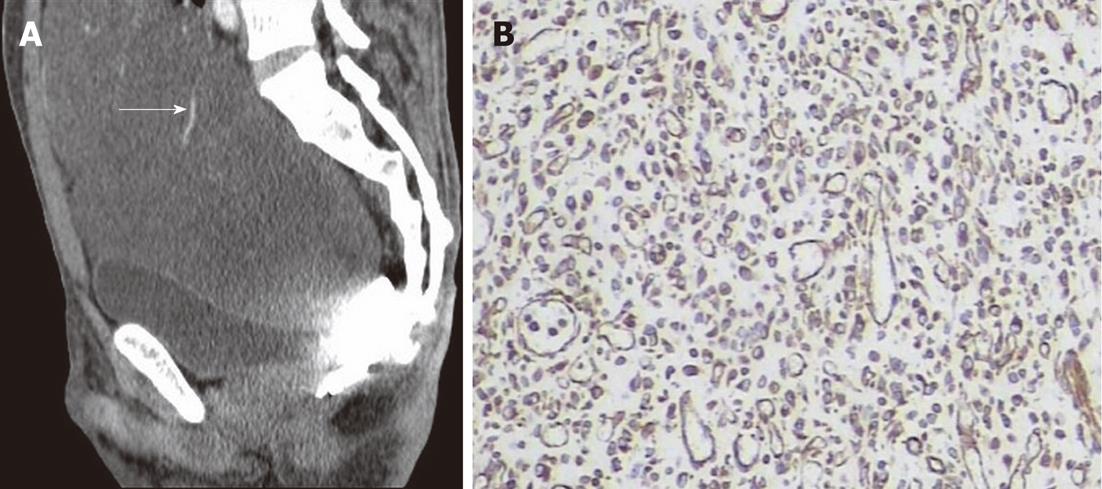
Figure 4 A 34-year-old male with greater omentum malignant fibrous histiocytoma.
A: Contrast-enhanced computed tomography scan sagittal view shows a well demarcated cystic mass extending from the abdominal to the pelvic cavity, note the blood vessel on the surface (arrow); B: Immunohistochemistry shows positive staining to vimentin (× 400).
- Citation: Karki B, Xu YK, Wu YK, Zhang WW. Primary malignant fibrous histiocytoma of the abdominal cavity: CT findings and pathological correlation. World J Radiol 2012; 4(4): 151-158
- URL: https://www.wjgnet.com/1949-8470/full/v4/i4/151.htm
- DOI: https://dx.doi.org/10.4329/wjr.v4.i4.151


