Copyright
©2012 Baishideng Publishing Group Co.
World J Radiol. Apr 28, 2012; 4(4): 141-150
Published online Apr 28, 2012. doi: 10.4329/wjr.v4.i4.141
Published online Apr 28, 2012. doi: 10.4329/wjr.v4.i4.141
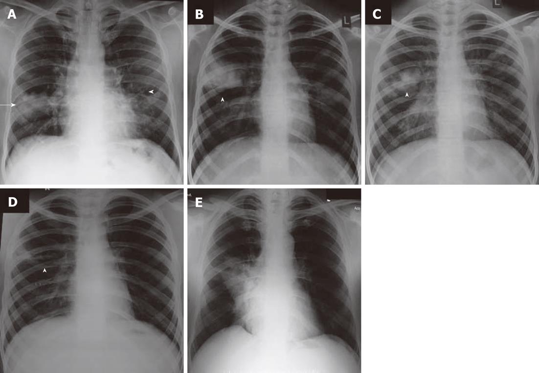
Figure 3 Chest radiograph.
A: Chest radiograph showing consolidation (arrow) and tram track opacities (arrowhead); B-D: Chest radiographs performed over time in a patient with allergic bronchopulmonary aspergillosis showing fleeting pulmonary opacities, and clearance after treatment. Arrowheads depict the fleeting opacities; E: Chest radiograph showing subsegmental collapse.
- Citation: Agarwal R, Khan A, Garg M, Aggarwal AN, Gupta D. Chest radiographic and computed tomographic manifestations in allergic bronchopulmonary aspergillosis. World J Radiol 2012; 4(4): 141-150
- URL: https://www.wjgnet.com/1949-8470/full/v4/i4/141.htm
- DOI: https://dx.doi.org/10.4329/wjr.v4.i4.141


