Copyright
©2012 Baishideng Publishing Group Co.
World J Radiol. Apr 28, 2012; 4(4): 128-134
Published online Apr 28, 2012. doi: 10.4329/wjr.v4.i4.128
Published online Apr 28, 2012. doi: 10.4329/wjr.v4.i4.128
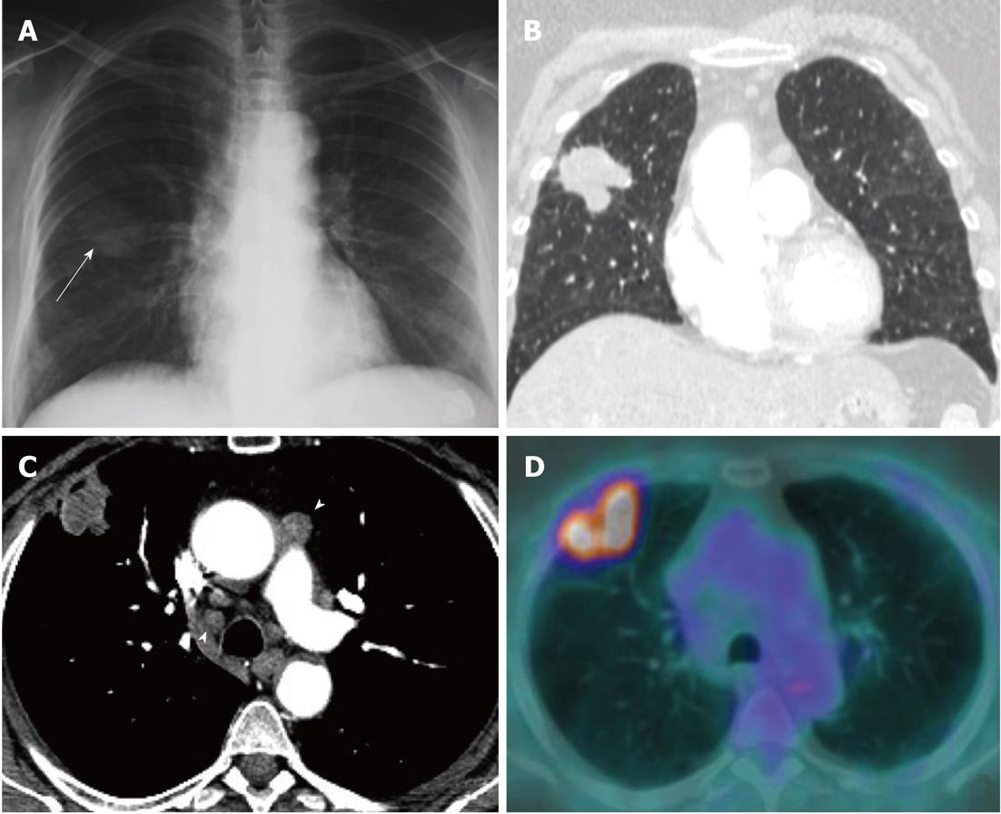
Figure 3 Example of cross sectional imaging nodal staging pitfalls.
A: A mass of the right lung (arrow) was identified on chest radiograph; B: A 4 cm spiculated mass suspected of lung cancer in the right upper lobe was confirmed by computed tomography (CT); C, D: Contrast enhanced CT demonstrated enlarged lymph nodes (> 1 cm in short axis; arrowheads) in ipsi- and contra-lateral mediastinal nodal stations (C) (T2aN3), positron emission tomography-computed tomography (PET-CT) (D) showed high metabolic activity of the parenchymal lesion but no nodal [18F]-2-fluoro-2-deoxy-d-glucose uptake (PET-CT staging: T2aN0M0). No metastatic nodes were demonstrated by endoscopic ultrasound-needle aspiration and mediastinoscopy, and surgical staging was in full agreement with PET-CT (pT2N0 adenocarcinoma).
- Citation: Mirsadraee S, Oswal D, Alizadeh Y, Caulo A, Beek EJV. The 7th lung cancer TNM classification and staging system: Review of the changes and implications. World J Radiol 2012; 4(4): 128-134
- URL: https://www.wjgnet.com/1949-8470/full/v4/i4/128.htm
- DOI: https://dx.doi.org/10.4329/wjr.v4.i4.128


