Copyright
©2012 Baishideng Publishing Group Co.
World J Radiol. Apr 28, 2012; 4(4): 128-134
Published online Apr 28, 2012. doi: 10.4329/wjr.v4.i4.128
Published online Apr 28, 2012. doi: 10.4329/wjr.v4.i4.128
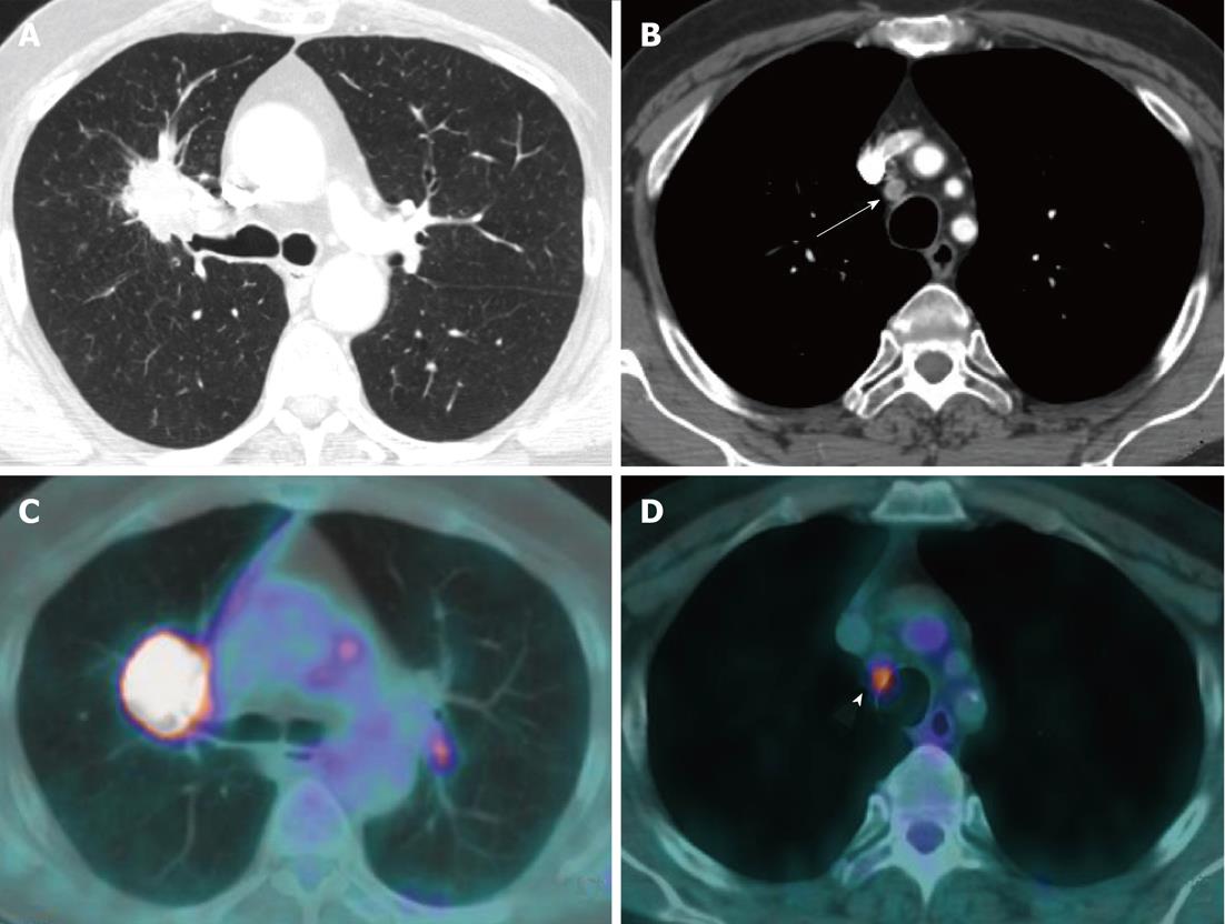
Figure 2 Contrast enhanced thoracic computed tomography viewed with lung window settings shows a 5.
2 cm ill-defined mass suspected of lung cancer abutting the right lung hilum, causing narrowing of the upper lobe bronchus (A). B: Review of images on mediastinal window settings showed a paratracheal lymph node with short axis smaller than 1 cm (arrow) in station 2R; C and D: Positron emission tomography-computed tomography demonstrated a high [18F]-2-fluoro-2-deoxy-d-glucose-uptake of both mass and 2R node (arrowhead), resulting in an up-staging of the disease from T2bN1 (CT classification) to T2bN2. This finding was further confirmed by mediastinoscopy sampling.
- Citation: Mirsadraee S, Oswal D, Alizadeh Y, Caulo A, Beek EJV. The 7th lung cancer TNM classification and staging system: Review of the changes and implications. World J Radiol 2012; 4(4): 128-134
- URL: https://www.wjgnet.com/1949-8470/full/v4/i4/128.htm
- DOI: https://dx.doi.org/10.4329/wjr.v4.i4.128


