Copyright
©2012 Baishideng Publishing Group Co.
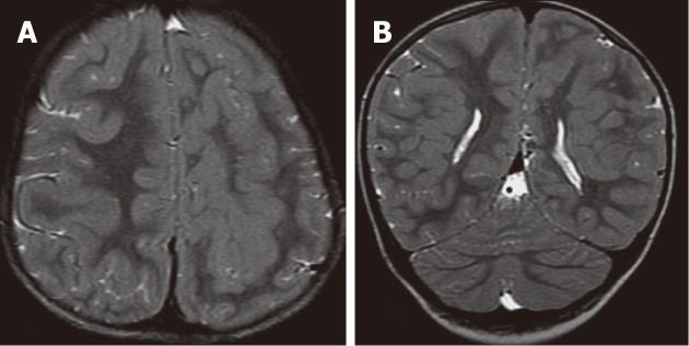
Figure 3 Subcortical curvilinear heterotopia.
A, B: Axial T2W and coronal T2W images showing bilateral curvilinear heterotopia within the white matter. The heterotopic tissue is convoluted and contiguous with the overlying cortex. Linear and punctuate cerebrospinal fluid signal are seen within the heterotopic tissue. The cerebral cortex shows pachygyria.
- Citation: Donkol RH, Moghazy KM, Abolenin A. Assessment of gray matter heterotopia by magnetic resonance imaging. World J Radiol 2012; 4(3): 90-96
- URL: https://www.wjgnet.com/1949-8470/full/v4/i3/90.htm
- DOI: https://dx.doi.org/10.4329/wjr.v4.i3.90


