Copyright
©2012 Baishideng Publishing Group Co.
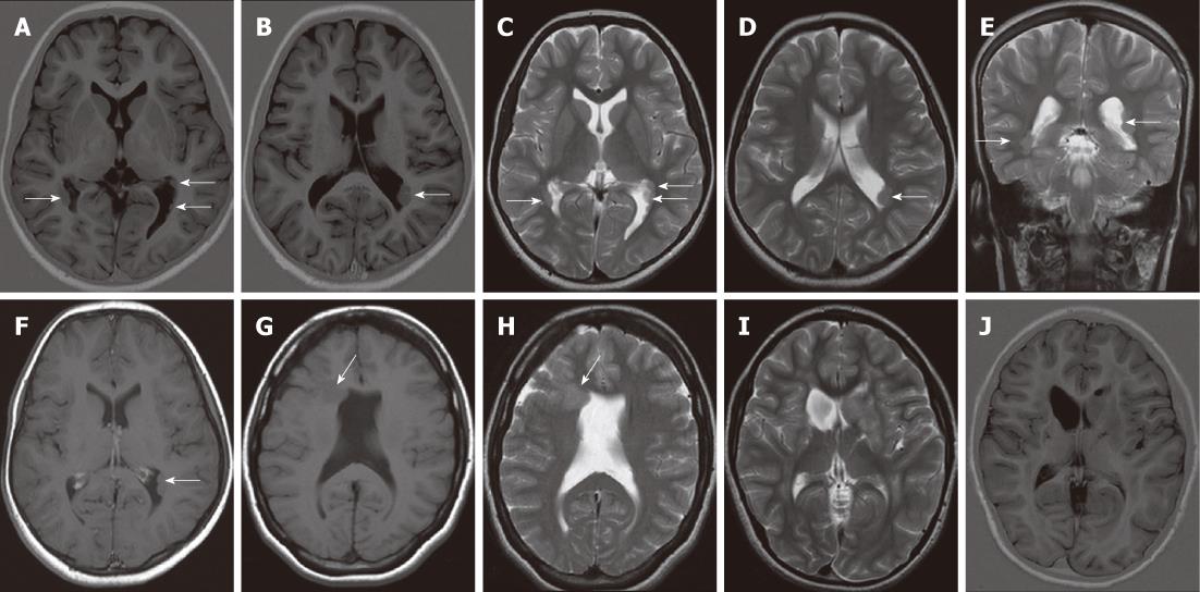
Figure 1 Subependymal heterotopia.
A, B: Axial inversion-recovery magnetic resonance (MR) images; C-E: Axial (C, D) and coronal T2 W (E) images showing multiple bilateral subependymal gray matter heterotopic nodules protruding into and indenting the trigone of the lateral ventricles and the left occipital horn (arrows). The nodules are isointense to cortical gray matter; F: Axial contrast-enhanced T1WI shows no enhancement of the nodules; G, H: Axial T1W and T2W MR images revealing unilateral focal subependymal gray matter nodule (arrow) protruding into and indenting the frontal horn of the right lateral ventricle. The nodule is isointense to the cortical gray matter. Mild ventricular dilatation is noted with absent septum pelucidum; I, J: Axial T2 WI and inversion-recovery MR image showing a unilateral, large subependymal heterotopic gray matter mass projecting into the frontal horn of the left lateral ventricle. The mass is isointense to the cortical gray matter.
- Citation: Donkol RH, Moghazy KM, Abolenin A. Assessment of gray matter heterotopia by magnetic resonance imaging. World J Radiol 2012; 4(3): 90-96
- URL: https://www.wjgnet.com/1949-8470/full/v4/i3/90.htm
- DOI: https://dx.doi.org/10.4329/wjr.v4.i3.90


