Copyright
©2012 Baishideng Publishing Group Co.
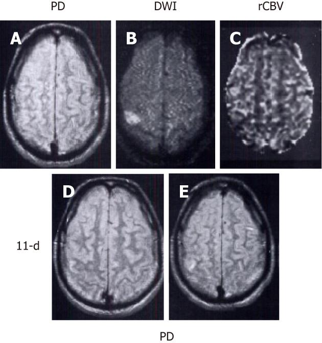
Figure 4 Main pattern of perfusion-diffusion mismatch: perfusion-weighted imaging < diffusion-weighted imaging, in a patient with acute stroke.
Patient’s left-sided weakness was partially resolved 3 h after the onset of symptoms. A: Proton density (PD) weighted fast spin-echo image shows no abnormality; B: Diffusion-weighted imaging (DWI) shows a focal cortical ischemic abnormality; C: Relative cerebral blood volume (rCBV) map demonstrates a smaller lesion with decreased rCBV compared to the same abnormality as depicted in B; D, E: Follow-up PD images acquired 11 d later show a new tiny hyperintense infarct in the area of initially observed lesion on B and C. Note that the initial DWI lesion is larger than the final infarct volume. The patient’s symptoms resolved completely after 2 d (reprint from Sorensen et al Radiology 1996; 199: 391-401 with permission).
- Citation: Chen F, Ni YC. Magnetic resonance diffusion-perfusion mismatch in acute ischemic stroke: An update. World J Radiol 2012; 4(3): 63-74
- URL: https://www.wjgnet.com/1949-8470/full/v4/i3/63.htm
- DOI: https://dx.doi.org/10.4329/wjr.v4.i3.63


