Copyright
©2012 Baishideng Publishing Group Co.
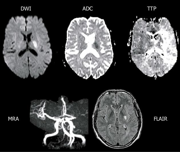
Figure 2 Main pattern of perfusion-diffusion mismatch, perfusion-weighted imaging > diffusion-weighted imaging, in a patient with acute stroke.
Extensive area of prolonged time to peak (TTP) and small diffusion-weighted imaging (DWI) lesion in deep middle cerebral artery (MCA) territory, with a complete proximal MCA occlusion on magnetic resonance angiography (MRA) (reprint from Muir KW et al Lancet Neurol 2006; 5: 755-68 with permission). ADC: Apparent diffusion coefficient; FLAIR: Fluid-attenuated inversion-recovery.
- Citation: Chen F, Ni YC. Magnetic resonance diffusion-perfusion mismatch in acute ischemic stroke: An update. World J Radiol 2012; 4(3): 63-74
- URL: https://www.wjgnet.com/1949-8470/full/v4/i3/63.htm
- DOI: https://dx.doi.org/10.4329/wjr.v4.i3.63


