Copyright
©2012 Baishideng Publishing Group Co.
World J Radiol. Mar 28, 2012; 4(3): 121-125
Published online Mar 28, 2012. doi: 10.4329/wjr.v4.i3.121
Published online Mar 28, 2012. doi: 10.4329/wjr.v4.i3.121
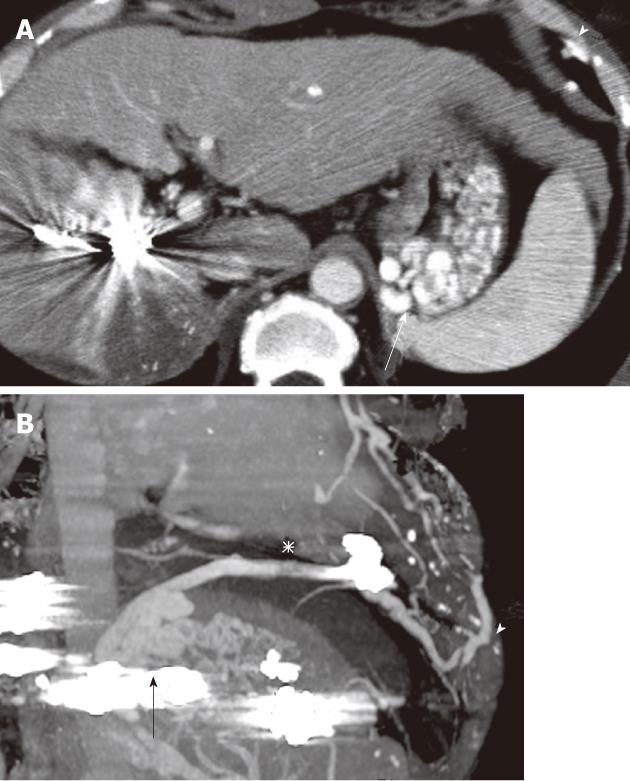
Figure 3 Portal phase of dynamic computed tomography images using contrast medium before balloon-occluded retrograde transvenous obliteration.
A: Axial image demonstrated enlargement of gastric varices (arrow) and intercostal vein (arrowhead) with marked shrinkage of hepatocellular carcinoma; B: Multi-planar reconstruction computed tomography image demonstrated the gastric varices (arrow) and the dilated phrenic branch (asterisk) communicating with intercostal vein (arrowhead).
-
Citation: Minamiguchi H, Kawai N, Sato M, Ikoma A, Sawa M, Sonomura T, Sahara S, Nakata K, Takasaka I, Nakai M. Balloon-occluded retrograde transvenous obliteration for gastric varices
via the intercostal vein. World J Radiol 2012; 4(3): 121-125 - URL: https://www.wjgnet.com/1949-8470/full/v4/i3/121.htm
- DOI: https://dx.doi.org/10.4329/wjr.v4.i3.121


