Copyright
©2012 Baishideng Publishing Group Co.
World J Radiol. Mar 28, 2012; 4(3): 121-125
Published online Mar 28, 2012. doi: 10.4329/wjr.v4.i3.121
Published online Mar 28, 2012. doi: 10.4329/wjr.v4.i3.121
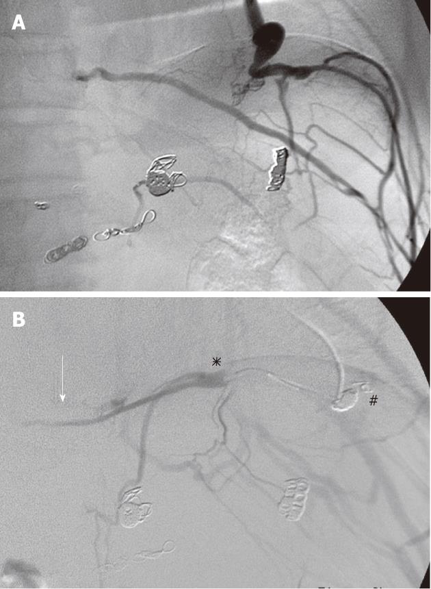
Figure 2 Micro-balloon-occluded pericardial venography.
A: Micro-balloon occluded venography (M-BOV) of the pericardial vein demonstrated the development of the cardio-phrenic vein and phrenic branch veins; B: Embolization of the cardio-phrenic vein using 9 microcoils (#) was conducted in order to reduce the collateral vessels. However, M-BOV of the left subphrenic vein (asterisk) followed by coil embolization demonstrated the narrow outlet of the subphrenic vein (arrow) and the dilated phrenic branches but not gastric varices. Further advancement of the micro-balloon catheter was difficult.
-
Citation: Minamiguchi H, Kawai N, Sato M, Ikoma A, Sawa M, Sonomura T, Sahara S, Nakata K, Takasaka I, Nakai M. Balloon-occluded retrograde transvenous obliteration for gastric varices
via the intercostal vein. World J Radiol 2012; 4(3): 121-125 - URL: https://www.wjgnet.com/1949-8470/full/v4/i3/121.htm
- DOI: https://dx.doi.org/10.4329/wjr.v4.i3.121


