Copyright
©2012 Baishideng Publishing Group Co.
World J Radiol. Mar 28, 2012; 4(3): 121-125
Published online Mar 28, 2012. doi: 10.4329/wjr.v4.i3.121
Published online Mar 28, 2012. doi: 10.4329/wjr.v4.i3.121
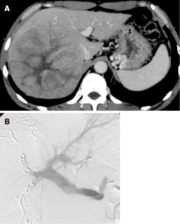
Figure 1 A 55-year-old man with gastric varices due to hepatitis B liver cirrhosis.
A: Hepatocellular carcinoma of 13 cm in the right lobe. Gastric varices were also demonstrated (arrow); B: Percutaneous transhepatic obliteration of the posterior gastric vein using 5% ethanolamine oleate-iopamidol and microcoils was performed to occlude the gastric varices. Percutaneous transhepatic portal vein embolization of the right posterior and anterior segments of the portal vein using lipiodol, gelatin sponge particles and microcoils was conducted using the same transhepatic catheter route in order to enlarge the left hepatic lobe for possible right hepatectomy.
-
Citation: Minamiguchi H, Kawai N, Sato M, Ikoma A, Sawa M, Sonomura T, Sahara S, Nakata K, Takasaka I, Nakai M. Balloon-occluded retrograde transvenous obliteration for gastric varices
via the intercostal vein. World J Radiol 2012; 4(3): 121-125 - URL: https://www.wjgnet.com/1949-8470/full/v4/i3/121.htm
- DOI: https://dx.doi.org/10.4329/wjr.v4.i3.121


