Copyright
©2012 Baishideng Publishing Group Co.
World J Radiol. Mar 28, 2012; 4(3): 115-120
Published online Mar 28, 2012. doi: 10.4329/wjr.v4.i3.115
Published online Mar 28, 2012. doi: 10.4329/wjr.v4.i3.115
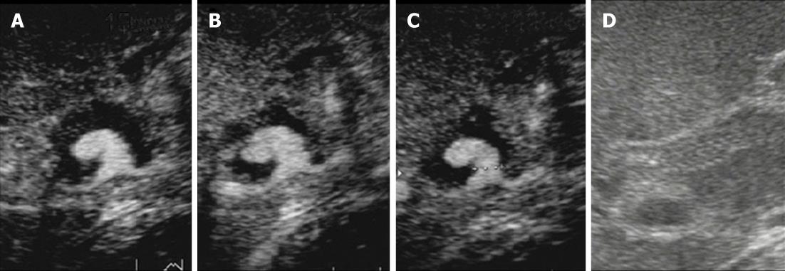
Figure 5 The pseudoaneurysm and hypoechoic solid components filling the dilated bile duct were examined on contrast-enhanced ultrasonography.
A-C: Images obtained at 15 s (A), 55 s (B) and 127 s (C) after injection of Sonazoid (0.5 mL) via a left cubital venous line showed no Sonazoid bubbles in the common bile duct other than in the pseudoaneurysm; D: Monitor B-mode ultrasonography image.
- Citation: Watanabe M, Shiozawa K, Mimura T, Ito K, Kamata I, Kishimoto Y, Momiyama K, Igarashi Y, Sumino Y. Hepatic artery pseudoaneurysm after endoscopic biliary stenting for bile duct cancer. World J Radiol 2012; 4(3): 115-120
- URL: https://www.wjgnet.com/1949-8470/full/v4/i3/115.htm
- DOI: https://dx.doi.org/10.4329/wjr.v4.i3.115


