Copyright
©2012 Baishideng Publishing Group Co.
World J Radiol. Mar 28, 2012; 4(3): 115-120
Published online Mar 28, 2012. doi: 10.4329/wjr.v4.i3.115
Published online Mar 28, 2012. doi: 10.4329/wjr.v4.i3.115
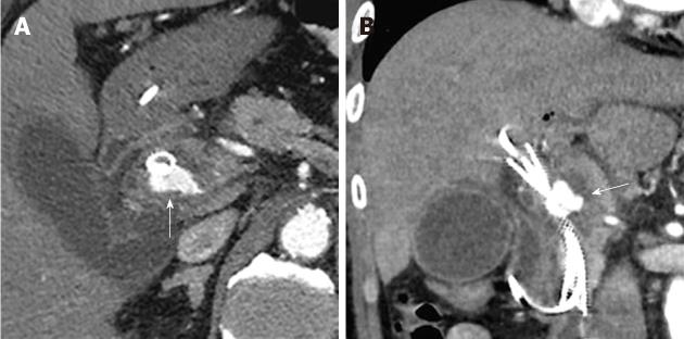
Figure 3 On the 2nd hospital day, an endoscopic naso-biliary drainage and a plastic stent were placed into the self-expandable metallic stent.
On the 6th hospital day, axial (A) and coronal (B) plane arterial phase computed tomography showed enlargement of the pseudoaneurysm (arrow).
- Citation: Watanabe M, Shiozawa K, Mimura T, Ito K, Kamata I, Kishimoto Y, Momiyama K, Igarashi Y, Sumino Y. Hepatic artery pseudoaneurysm after endoscopic biliary stenting for bile duct cancer. World J Radiol 2012; 4(3): 115-120
- URL: https://www.wjgnet.com/1949-8470/full/v4/i3/115.htm
- DOI: https://dx.doi.org/10.4329/wjr.v4.i3.115


