Copyright
©2012 Baishideng Publishing Group Co.
World J Radiol. Mar 28, 2012; 4(3): 109-114
Published online Mar 28, 2012. doi: 10.4329/wjr.v4.i3.109
Published online Mar 28, 2012. doi: 10.4329/wjr.v4.i3.109
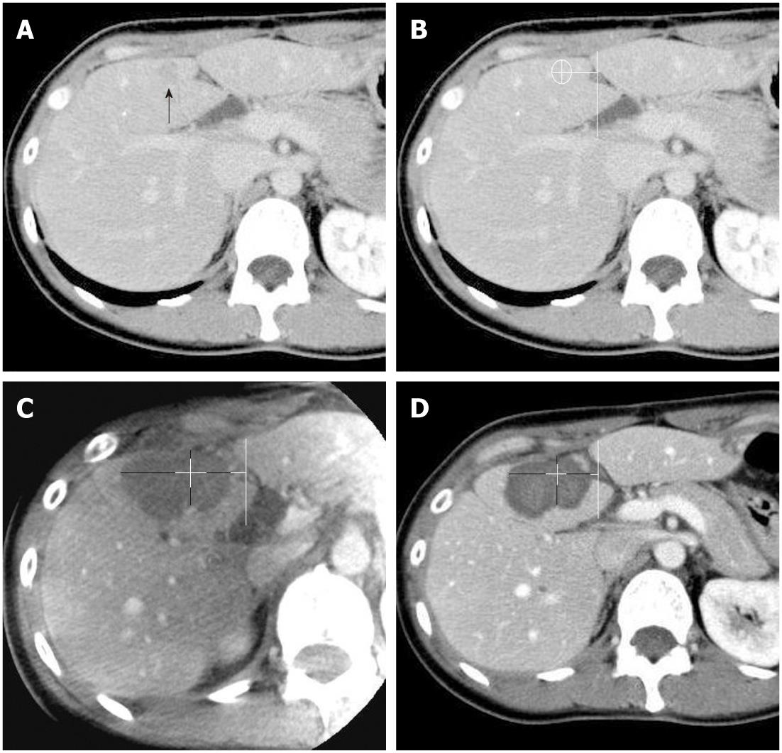
Figure 1 A 43-year-old woman with a 13-mm hepatocellular carcinoma in hepatic segment IV.
A: A venous-phase image from contrast-enhanced multidetector computed tomography (MDCT) prior to treatment shows a hypoattenuated tumor in hepatic segment IV (arrow); B: A venous-phase MDCT image in which the tumor size has been measured in vertical and horizontal directions and the tumor location has been determined by the distance from the left portal vein; C: An intravenous contrast-enhanced C-arm computed tomography image obtained just after radiofrequency ablation (RFA) shows sufficient ablative margins in all 4 directions (ventral margin, 1.8 mm; dorsal margin, 8.8 mm; right lateral margin, 20.5 mm, left lateral margin, 10.3 mm) even though the ventral ablative margin is less than 5 mm due to the adjacent liver border; D: A portal-phase image from contrast-enhanced MDCT obtained 7 d after the RFA procedure shows an almost identical configuration with ablative margins (ventral margin, 2.0 mm; dorsal margin, 8.0 mm; right lateral margin, 17.0 mm; left lateral margin, 10.0 mm) comparable to those depicted in the C-arm CT image.
- Citation: Iwazawa J, Ohue S, Hashimoto N, Mitani T. Ablation margin assessment of liver tumors with intravenous contrast-enhanced C-arm computed tomography. World J Radiol 2012; 4(3): 109-114
- URL: https://www.wjgnet.com/1949-8470/full/v4/i3/109.htm
- DOI: https://dx.doi.org/10.4329/wjr.v4.i3.109


