Copyright
©2012 Baishideng Publishing Group Co.
World J Radiol. Mar 28, 2012; 4(3): 102-108
Published online Mar 28, 2012. doi: 10.4329/wjr.v4.i3.102
Published online Mar 28, 2012. doi: 10.4329/wjr.v4.i3.102
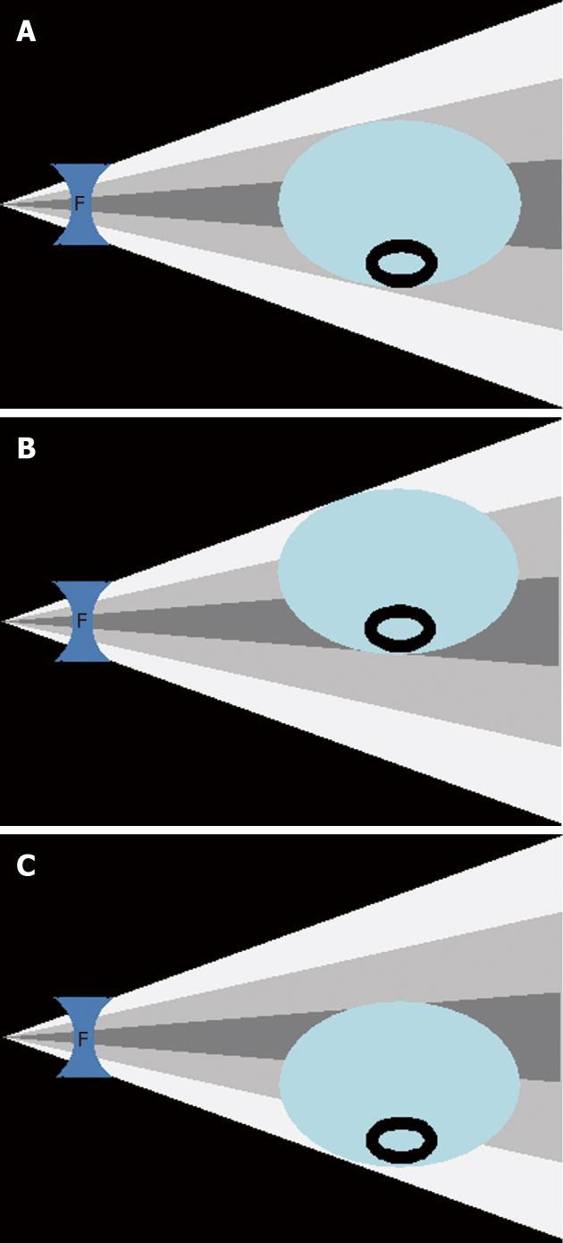
Figure 2 More attenuated X-ray beam is received in the center of bow-tie filter corresponds to the posterior abdominal wall and the anterior abdominal wall when patients are centered the gantry isocenter.
A: When the patient is centered in the gantry isocenter, bow-fie filter (F) allows most X-rays (darker gray) to traverse thicker, central parts of the patient and fewer X-rays (white) in the peripheral portion to pass through thinner, peripheral parts of the patient; B: When the patient is positioned above the gantry isocenter, the anterior portion of the patient’s abdominal wall which is further from the gantry isocenter receives fewer X-rays (white) with a consequent increase in noise, whereas the posterior abdominal wall receives the central more intense X-ray beam (darker gray), and therefore has less noise; C: When the patient is positioned below the gantry isocenter, the anterior portion of the patient’s abdominal wall coincides with the gantry isocenter to receive more X-rays, resulting in no significant change in its noise, whereas the posterior abdominal wall moves away from the gantry isocenter and receives fewer X-rays which leads to higher noise.
- Citation: Kim MS, Singh S, Halpern E, Saini S, Kalra MK. Ablation margin assessment of liver tumors with intravenous contrast-enhanced C-arm computed tomography. World J Radiol 2012; 4(3): 102-108
- URL: https://www.wjgnet.com/1949-8470/full/v4/i3/102.htm
- DOI: https://dx.doi.org/10.4329/wjr.v4.i3.102


