Copyright
©2012 Baishideng Publishing Group Co.
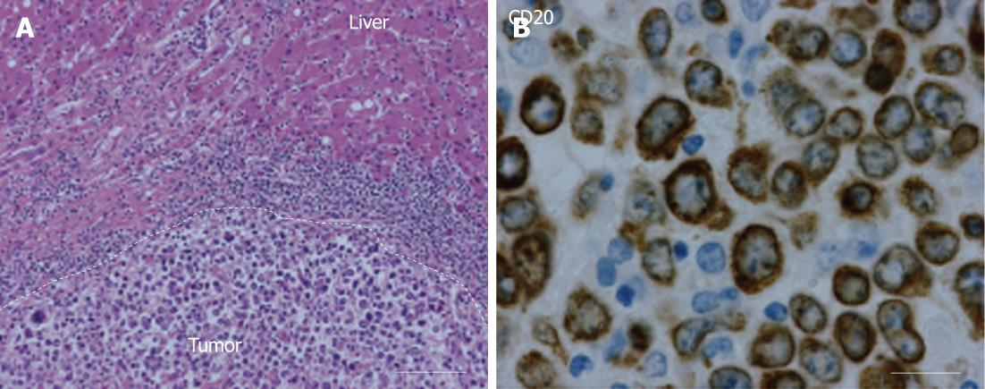
Figure 4 Histopathological results.
A: Hematoxylin eosin staining (150 ×) shows a large cell malignancy with mostly loose tumor cells with nuclear polymorphism. Furthermore, frequent giant nuclear bodies with macro-nucleoli and numerous cell mitoses. No central necrosis is observed; B: A photomicrograph (400 ×) showing lymphocytic tumor cells which are positive for CD20 staining around the plasma membrane, indicating non-Hodgkin lymphoma of B-cell origin.
- Citation: Steller EJ, Leeuwen MSV, Hillegersberg RV, Schipper ME, Rinkes IHB, Molenaar IQ. Primary lymphoma of the liver - A complex diagnosis. World J Radiol 2012; 4(2): 53-57
- URL: https://www.wjgnet.com/1949-8470/full/v4/i2/53.htm
- DOI: https://dx.doi.org/10.4329/wjr.v4.i2.53


