Copyright
©2012 Baishideng Publishing Group Co.
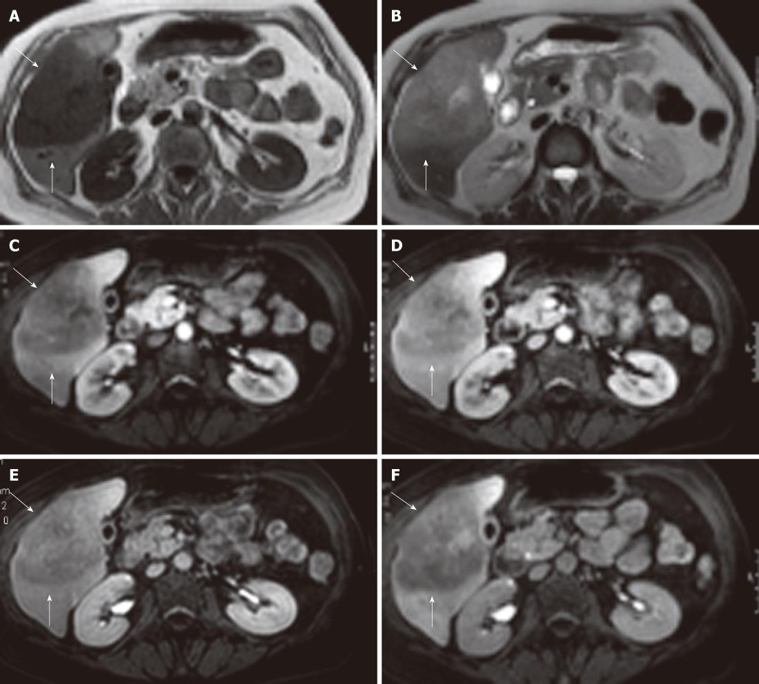
Figure 2 Magnetic resonance imaging showing a large, sharply demarcated lesion measuring 11 cm in the right liver lobe.
The lesion is hypointense on T1 (A) and hyperintense on T2 (B) weighed images with slight inhomogeneity. Arterial (C), portal (D), equilibrium (E) and hepato-biliary (20 min) (F) phase magnetic resonance imaging after Gd-EOB-DTPA contrast enhancement reveal a hypovasular lesion without uptake in the hepato-biliary phase (lesion indicated by white arrows).
- Citation: Steller EJ, Leeuwen MSV, Hillegersberg RV, Schipper ME, Rinkes IHB, Molenaar IQ. Primary lymphoma of the liver - A complex diagnosis. World J Radiol 2012; 4(2): 53-57
- URL: https://www.wjgnet.com/1949-8470/full/v4/i2/53.htm
- DOI: https://dx.doi.org/10.4329/wjr.v4.i2.53


