Copyright
©2012 Baishideng Publishing Group Co.
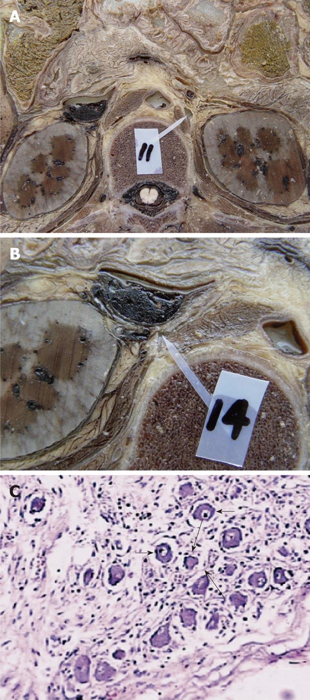
Figure 5 The dissection of the celiac ganglia in a cadaver at the L1 level.
A: The left celiac ganglia; B: The right celiac ganglia; C: The histologic specimen stained with hematoxylin-eosin staining (× 100). With light microscopy, the celiac ganglion shows scattered ganglion cells (short arrows) and sparse nerve fibers (long arrows) among these ganglion cells.
- Citation: Zuo HD, Zhang XM, Li CJ, Cai CP, Zhao QH, Xie XG, Xiao B, Tang W. CT and MR imaging patterns for pancreatic carcinoma invading the extrapancreatic neural plexus (Part I): Anatomy, imaging of the extrapancreatic nerve. World J Radiol 2012; 4(2): 36-43
- URL: https://www.wjgnet.com/1949-8470/full/v4/i2/36.htm
- DOI: https://dx.doi.org/10.4329/wjr.v4.i2.36


