Copyright
©2012 Baishideng Publishing Group Co.
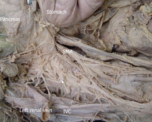
Figure 4 The anatomy of the celiac ganglia in a cadaver.
The celiac ganglion (white arrows) was close to the aorta at a level intermediate to the origin of the celiac artery and superior to the mesenteric artery. It was located in the space bound by the inferior vena cava (IVC), the head of pancreas and the superior mesenteric artery (SMA). The pancreas was moved upward. The IVC was cut and moved laterally.
- Citation: Zuo HD, Zhang XM, Li CJ, Cai CP, Zhao QH, Xie XG, Xiao B, Tang W. CT and MR imaging patterns for pancreatic carcinoma invading the extrapancreatic neural plexus (Part I): Anatomy, imaging of the extrapancreatic nerve. World J Radiol 2012; 4(2): 36-43
- URL: https://www.wjgnet.com/1949-8470/full/v4/i2/36.htm
- DOI: https://dx.doi.org/10.4329/wjr.v4.i2.36


