Copyright
©2012 Baishideng Publishing Group Co.
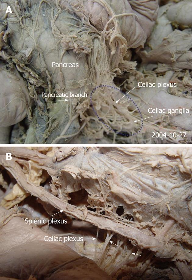
Figure 3 The anatomy of the celiac plexus in cadavers (A, B).
The pancreas was moved upward. The pictures demonstrate that the celiac plexus is the center of the viscus and composed of the celiac ganglia and several large and small nerves that terminate with the celiac ganglia (arrows).
- Citation: Zuo HD, Zhang XM, Li CJ, Cai CP, Zhao QH, Xie XG, Xiao B, Tang W. CT and MR imaging patterns for pancreatic carcinoma invading the extrapancreatic neural plexus (Part I): Anatomy, imaging of the extrapancreatic nerve. World J Radiol 2012; 4(2): 36-43
- URL: https://www.wjgnet.com/1949-8470/full/v4/i2/36.htm
- DOI: https://dx.doi.org/10.4329/wjr.v4.i2.36


