Copyright
©2012 Baishideng Publishing Group Co.
World J Radiol. Nov 18, 2012; 4(11): 443-449
Published online Nov 18, 2012. doi: 10.4329/wjr.v4.i11.443
Published online Nov 18, 2012. doi: 10.4329/wjr.v4.i11.443
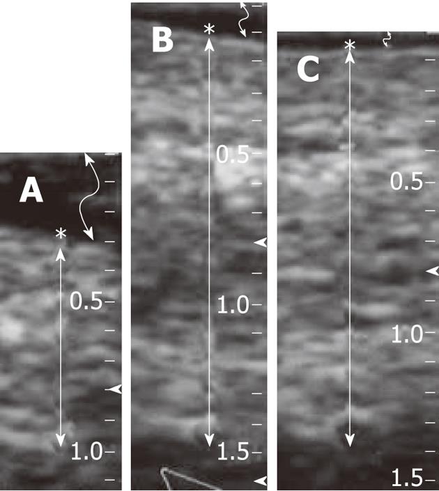
Figure 3 Measurement of skin thickness: The skin thickness from the epidermis to the bottom of the subdermis was approximately 7 mm before the gel injection (A), approx.
Fourteen mm after gel injection (B) and the thickness almost remained the same after irradiation (C). Slight decrease in the thickness might be due to compression by the probe. A low echogenic gel area is seen in the mid zone of the subcutaneous tissue including bright spots of air bubbles (B and C). Asterisk: Skin surface (epidermis); While arrow: Range of skin including epidermis, dermis and subdermis; Curved arrow: A gel layer between the surface of the skin and the ultrasonography probe (top), which surface gel was for a precise measurement avoiding compression.
- Citation: Kishi K, Tanino H, Sonomura T, Shirai S, Noda Y, Sato M, Okamura Y. Novel eradicative high-dose rate brachytherapy for internal mammary lymph node metastasis from breast cancer. World J Radiol 2012; 4(11): 443-449
- URL: https://www.wjgnet.com/1949-8470/full/v4/i11/443.htm
- DOI: https://dx.doi.org/10.4329/wjr.v4.i11.443


