Copyright
©2012 Baishideng Publishing Group Co.
World J Radiol. Nov 18, 2012; 4(11): 443-449
Published online Nov 18, 2012. doi: 10.4329/wjr.v4.i11.443
Published online Nov 18, 2012. doi: 10.4329/wjr.v4.i11.443
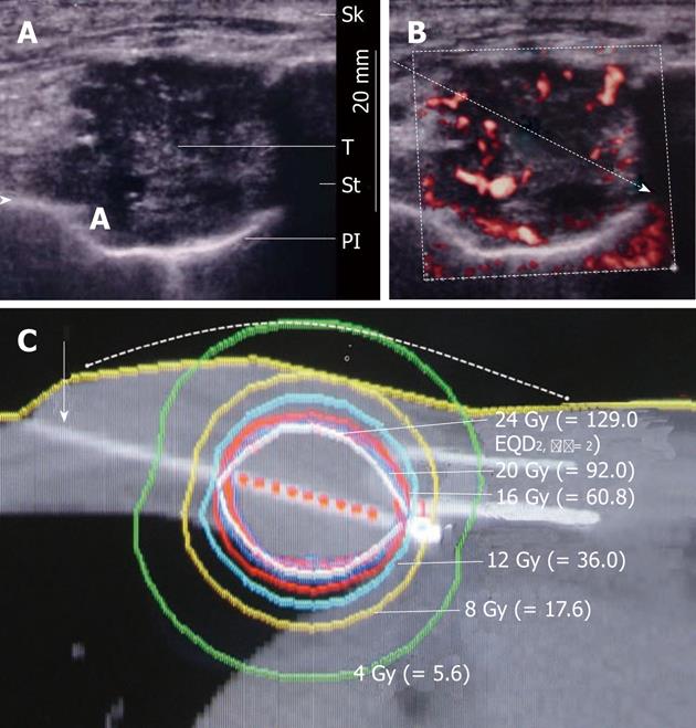
Figure 2 Plain ultrasound image (A) and power Doppler image showing the planned route for needle insertion to avoid vessels, pleura and lung (B): Brachytherapy dose distribution and inserted brachytherapy needle (white arrow) with nine red points (interval: 2.
5 mm) for source activation. The dashed line shows a supposed skin surface line raised by 7.0 mm and a single dot and a circle show the shifted dermal and subdermal reference point, respectively (C). The skin density is increased by subcutaneous injection of lidocaine, not by skin invasion. Dotted circle indicates planning target volume contour. Isodose curves are 150% (white), 125% (blue), 100% (red), 75% (light blue), 50% (yellow) and 25% (green) of 16 Gy, from innermost to outermost. Sk: Skin; T: Tumor; St: Sternum; Pl: Pleura.
- Citation: Kishi K, Tanino H, Sonomura T, Shirai S, Noda Y, Sato M, Okamura Y. Novel eradicative high-dose rate brachytherapy for internal mammary lymph node metastasis from breast cancer. World J Radiol 2012; 4(11): 443-449
- URL: https://www.wjgnet.com/1949-8470/full/v4/i11/443.htm
- DOI: https://dx.doi.org/10.4329/wjr.v4.i11.443


