Copyright
©2012 Baishideng Publishing Group Co.
World J Radiol. Oct 28, 2012; 4(10): 439-442
Published online Oct 28, 2012. doi: 10.4329/wjr.v4.i10.439
Published online Oct 28, 2012. doi: 10.4329/wjr.v4.i10.439
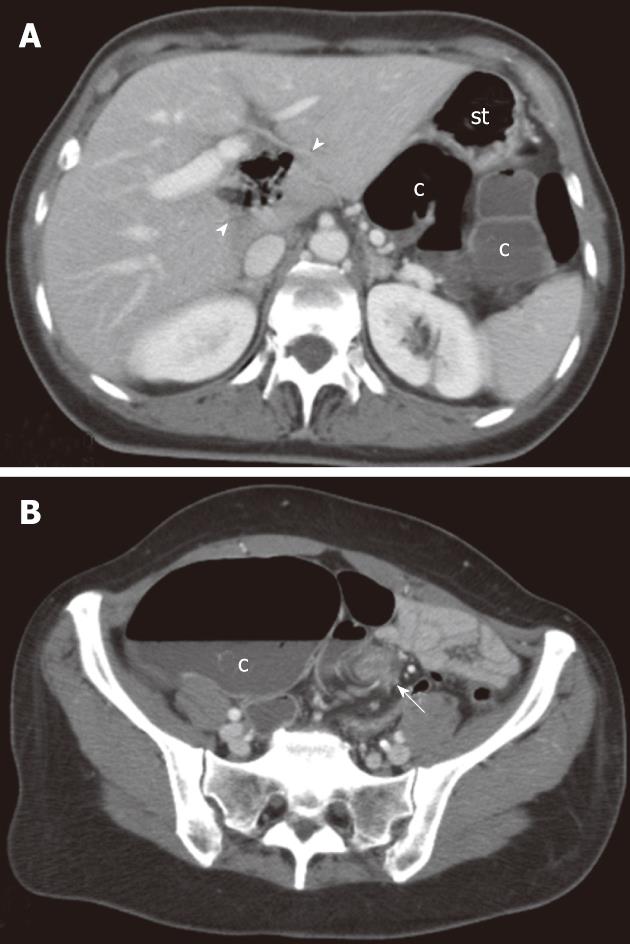
Figure 2 Multi-detector contrast-enhanced computed tomography.
Transverse images at the level of the upper abdomen (A) and the pelvis (B) are shown. A: A gas-containing loop is clearly depicted within the hepatic hilum (arrowheads) along with the evidence of a colonic segment (c) situated behind the stomach (st); B: An abnormally dilated colonic segment misinterpreted on the axial plane as cecum (c) can be appreciated in the right iliac fossa whereas torsion of the mesenteric vascular axis (whirl sign) is clearly depicted in the left iliac fossa (arrow).
- Citation: Camera L, Calabrese M, Mainenti PP, Masone S, Vecchio WD, Persico G, Salvatore M. Volvulus of the ascending colon in a non-rotated midgut: Plain film and MDCT findings. World J Radiol 2012; 4(10): 439-442
- URL: https://www.wjgnet.com/1949-8470/full/v4/i10/439.htm
- DOI: https://dx.doi.org/10.4329/wjr.v4.i10.439


