Copyright
©2012 Baishideng Publishing Group Co.
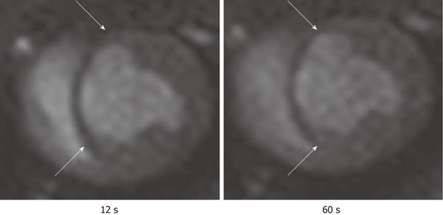
Figure 9 First pass perfusion magnetic resonance imaging shows the hypoperfused infarct scar (arrows) after bolus administration of Gd-DTPA.
The magnetic resonance (MR) images illustrate the delay in the enhancement of infarcted myocardium (arrows) compared with remote myocardium. The images were acquired at 12 s (left) and 60 s (right) after bolus administration of MR contrast media.
- Citation: Saeed M, Wilson M. Value of MR contrast media in image-guided body interventions. World J Radiol 2012; 4(1): 1-12
- URL: https://www.wjgnet.com/1949-8470/full/v4/i1/1.htm
- DOI: https://dx.doi.org/10.4329/wjr.v4.i1.1


