Copyright
©2012 Baishideng Publishing Group Co.
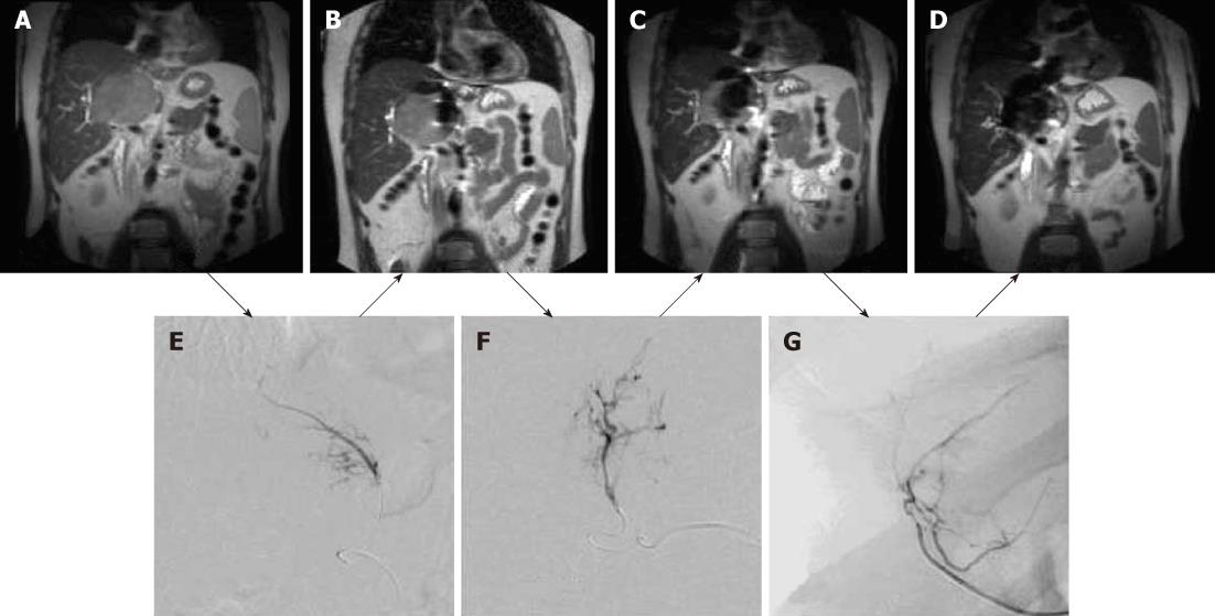
Figure 6 Coronal magnetic resonance images of large hepatocellular carcinoma obtained before magnetic targeted carrier bound to doxorubicin administration (A) and after the first (B), second (C) and third (D) dose of magnetic targeted carrier bound to doxorubicin.
The selective hepatic arterial catheter was repositioned between each dose. The initial DSA image (E) was obtained during the injection of magnetic targeted carrier bound to doxorubicin (MTC-DOX) into the left hepatic artery supplying the tumor. The next dose was injected into the hepatic artery branch segment (F), while the third dose was injected into a branch of the right hepatic artery (G). As a result, progressively larger areas of the tumor were affected by MTC-DOX, as documented by the progressive loss of signal intensity due to iron susceptibility artifacts (T2*) on the intra-procedural coronal magnetic resonance images obtained after each injection.
- Citation: Saeed M, Wilson M. Value of MR contrast media in image-guided body interventions. World J Radiol 2012; 4(1): 1-12
- URL: https://www.wjgnet.com/1949-8470/full/v4/i1/1.htm
- DOI: https://dx.doi.org/10.4329/wjr.v4.i1.1


