Copyright
©2012 Baishideng Publishing Group Co.
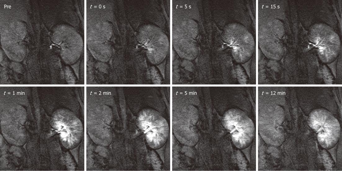
Figure 4 Distribution of magnetic resonance labeled embolic materials as a function of time in the left kidney.
Gadolinium-impregnated microspheres caused a steep increase in signal intensity over the cortical and medullary regions. Less than 5 min after the injection, as excess free gadolinium was excreted in the urine, the signal intensities began to decrease but still remained substantially above baseline for both particle sizes. Administration of magnetic resonance contrast media after the procedure confirmed the hypothesis that the injection of microspheres halts blood flow to targeted tissues. Hyperenhanced foci corresponding to microsphere location persisted for at least 1hr after injections. The smaller particles tended to settle more peripherally in the renal cortex, whereas the larger microspheres were lodged in the inner cortical and medullary regions. This new labeling technique may be useful for embolotherapy procedures, such as uterine fibroid embolization, hepatic tumor embolization, and preoperative meningioma embolization.
- Citation: Saeed M, Wilson M. Value of MR contrast media in image-guided body interventions. World J Radiol 2012; 4(1): 1-12
- URL: https://www.wjgnet.com/1949-8470/full/v4/i1/1.htm
- DOI: https://dx.doi.org/10.4329/wjr.v4.i1.1


