Copyright
©2012 Baishideng Publishing Group Co.
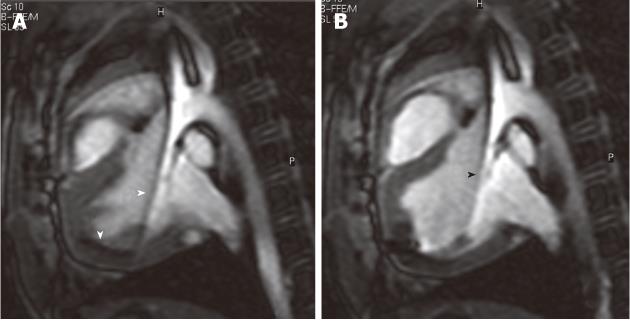
Figure 3 Selected magnetic resonance-guided images showing the advancement of a passive injecting catheter (arrowheads) in the left ventricle prior to (A) and after injecting dysprosium-chelate (T2* enhancing agent) magnetic resonance contrast medium (B, arrowhead) to ensure delivery in the targeted myocardium.
- Citation: Saeed M, Wilson M. Value of MR contrast media in image-guided body interventions. World J Radiol 2012; 4(1): 1-12
- URL: https://www.wjgnet.com/1949-8470/full/v4/i1/1.htm
- DOI: https://dx.doi.org/10.4329/wjr.v4.i1.1


