Copyright
©2011 Baishideng Publishing Group Co.
World J Radiol. Sep 28, 2011; 3(9): 224-232
Published online Sep 28, 2011. doi: 10.4329/wjr.v3.i9.224
Published online Sep 28, 2011. doi: 10.4329/wjr.v3.i9.224
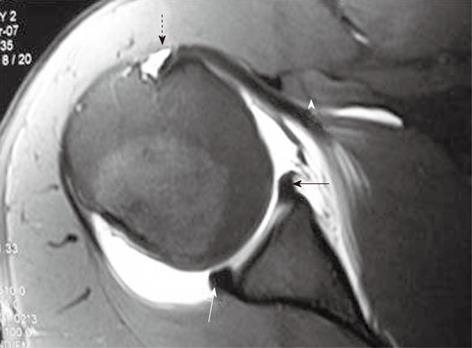
Figure 1 Normal T1-weighted TSE fat-saturated axial magnetic resonance arthrogram image.
The anterior and posterior labrum appears as triangular hypointense structures (straight arrows). Normal middle glenohumeral ligament has been shown with an arrowhead. Note the long head of biceps tendon in the bicipital groove and extension of joint fluid around the tendon (dashed arrow).
- Citation: Jana M, Gamanagatti S. Magnetic resonance imaging in glenohumeral instability. World J Radiol 2011; 3(9): 224-232
- URL: https://www.wjgnet.com/1949-8470/full/v3/i9/224.htm
- DOI: https://dx.doi.org/10.4329/wjr.v3.i9.224


