Copyright
©2011 Baishideng Publishing Group Co.
World J Radiol. Sep 28, 2011; 3(9): 215-223
Published online Sep 28, 2011. doi: 10.4329/wjr.v3.i9.215
Published online Sep 28, 2011. doi: 10.4329/wjr.v3.i9.215
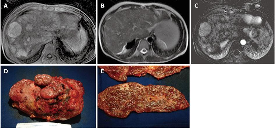
Figure 13 A 13-year-old male post-Kasai who underwent surgical spleno-renal shunt for gastrointestinal bleeding.
A: Axial T1 Fat Sat weighted acquisition shows multiple hyperintense nodules, the largest of these in S8; B: Axial T2 weighted acquisition: the nodules are isointense; C: Axial T1 Fat Sat weighted acquisition obtained with digital subtraction between the arterial phase and the unenhanced phase shows the vascular enhancement of the nodules; D: Explanted liver shows an irregular and multilobulated surface; E: Gross examination of sectioned liver confirms the presence of multiple regenerative nodular hyperplasia nodules.
- Citation: Miraglia R, Caruso S, Maruzzelli L, Spada M, Riva S, Sciveres M, Luca A. MDCT, MR and interventional radiology in biliary atresia candidates for liver transplantation. World J Radiol 2011; 3(9): 215-223
- URL: https://www.wjgnet.com/1949-8470/full/v3/i9/215.htm
- DOI: https://dx.doi.org/10.4329/wjr.v3.i9.215


