Copyright
©2011 Baishideng Publishing Group Co.
World J Radiol. Sep 28, 2011; 3(9): 215-223
Published online Sep 28, 2011. doi: 10.4329/wjr.v3.i9.215
Published online Sep 28, 2011. doi: 10.4329/wjr.v3.i9.215
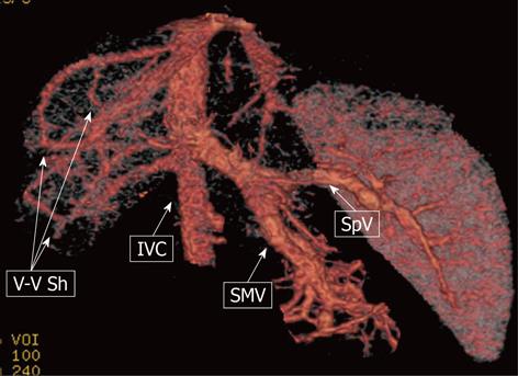
Figure 9 A 9-mo-old male child.
Multi-detector computed tomography: volume rendering reconstruction shows the superior mesenteric vein (SMV) and the splenic vein (SpV) joining to form a short common trunk that directly enters into the suprarenal inferior vena cava (IVC); Venous-venous intrahepatic shunting (V-V Sh) coexist.
- Citation: Miraglia R, Caruso S, Maruzzelli L, Spada M, Riva S, Sciveres M, Luca A. MDCT, MR and interventional radiology in biliary atresia candidates for liver transplantation. World J Radiol 2011; 3(9): 215-223
- URL: https://www.wjgnet.com/1949-8470/full/v3/i9/215.htm
- DOI: https://dx.doi.org/10.4329/wjr.v3.i9.215


