Copyright
©2011 Baishideng Publishing Group Co.
World J Radiol. Jul 28, 2011; 3(7): 194-198
Published online Jul 28, 2011. doi: 10.4329/wjr.v3.i7.194
Published online Jul 28, 2011. doi: 10.4329/wjr.v3.i7.194
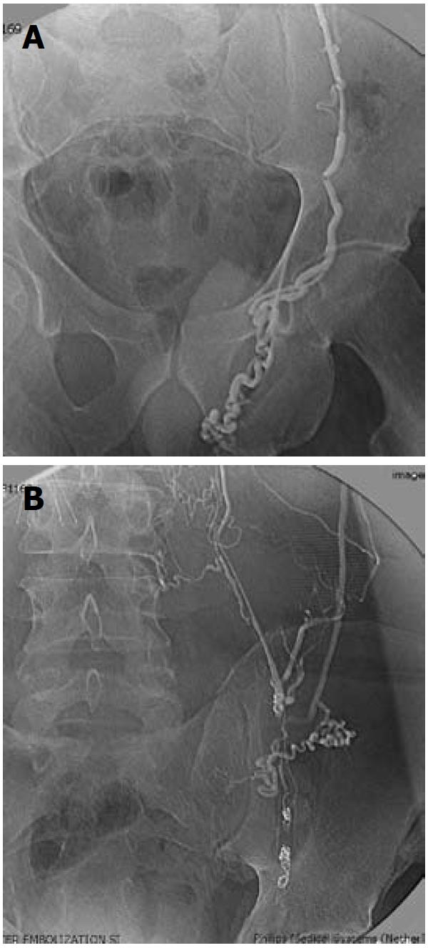
Figure 1 Venography following transjugular access.
A: Left gonadal venogram with transjugular access demonstrating large left varicocele; B: Left gonadal venogram, following coil occlusion of the gonadal vein at the internal inguinal ring, demonstrating extensive retroperitoneal collateralization.
- Citation: Gendel V, Haddadin I, Nosher JL. Antegrade pampiniform plexus venography in recurrent varicocele: Case report and anatomy review. World J Radiol 2011; 3(7): 194-198
- URL: https://www.wjgnet.com/1949-8470/full/v3/i7/194.htm
- DOI: https://dx.doi.org/10.4329/wjr.v3.i7.194


