Copyright
©2011 Baishideng Publishing Group Co.
World J Radiol. Jul 28, 2011; 3(7): 182-187
Published online Jul 28, 2011. doi: 10.4329/wjr.v3.i7.182
Published online Jul 28, 2011. doi: 10.4329/wjr.v3.i7.182
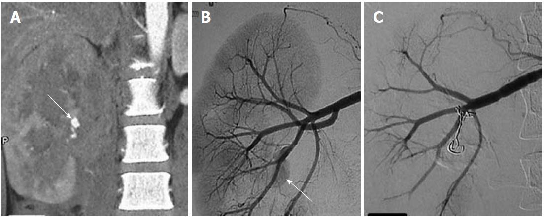
Figure 7 Traumatic right renal pseudoaneurysm.
A: Coronal maximum intensity projection computed tomography angiographic image reveals a large renal midpolar laceration and a pseudoaneurysm (arrow); B: Selective angiogram of the main right renal artery reveals a pseudoaneurysm (arrow) arising from one of the posterior branches; C: Successfully occluded using coil.
- Citation: Jana M, Gamanagatti S, Mukund A, Paul S, Gupta P, Garg P, Chattopadhyay TK, Sahni P. Endovascular management in abdominal visceral arterial aneurysms: A pictorial essay. World J Radiol 2011; 3(7): 182-187
- URL: https://www.wjgnet.com/1949-8470/full/v3/i7/182.htm
- DOI: https://dx.doi.org/10.4329/wjr.v3.i7.182


