Copyright
©2011 Baishideng Publishing Group Co.
World J Radiol. Jul 28, 2011; 3(7): 182-187
Published online Jul 28, 2011. doi: 10.4329/wjr.v3.i7.182
Published online Jul 28, 2011. doi: 10.4329/wjr.v3.i7.182
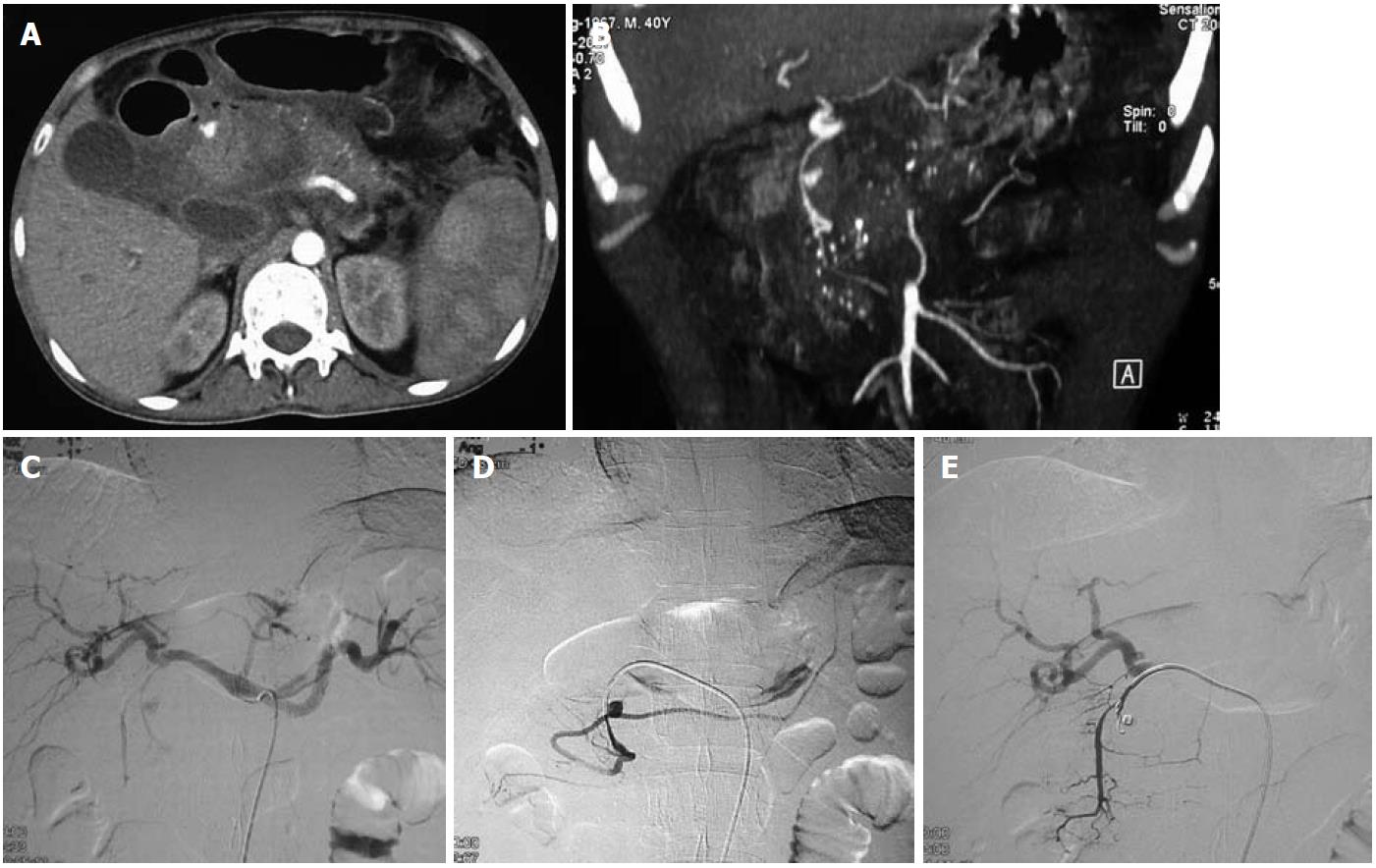
Figure 5 Endovascular management of a gastroduodenal artery aneurysm secondary to chronic calcific pancreatitis.
A, B: Axial image (A) and coronal reformatted image (B) of a computed tomography angiogram reveal features of acute on chronic calcific pancreatitis and a small saccular pseudoaneurysm of the gastroduodenal artery; C, D: Selective angiogram of the celiac axis (C) and gastroduodenal artery (D) reveal the filling of the aneurysm from the main trunk; E: Coil embolization was performed to fill the aneurysm cavity and cause complete occlusion.
- Citation: Jana M, Gamanagatti S, Mukund A, Paul S, Gupta P, Garg P, Chattopadhyay TK, Sahni P. Endovascular management in abdominal visceral arterial aneurysms: A pictorial essay. World J Radiol 2011; 3(7): 182-187
- URL: https://www.wjgnet.com/1949-8470/full/v3/i7/182.htm
- DOI: https://dx.doi.org/10.4329/wjr.v3.i7.182


