Copyright
©2011 Baishideng Publishing Group Co.
World J Radiol. Jul 28, 2011; 3(7): 182-187
Published online Jul 28, 2011. doi: 10.4329/wjr.v3.i7.182
Published online Jul 28, 2011. doi: 10.4329/wjr.v3.i7.182
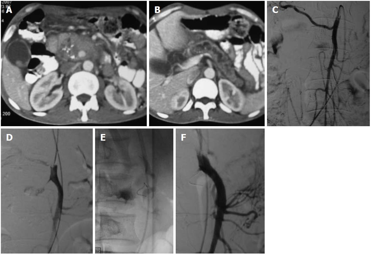
Figure 4 Coil embolization in an superior mesenteric artery aneurysm in a patient with chronic calcific pancreatitis.
A, B: Axial contrast-enhanced computed tomography of the abdomen revealed dilated main pancreatic duct and coarse calcification in the head of the pancreas, a large partially thrombosed pseudoaneurysm was apparent as a contrast filled globular structure in the head; C, D: Selective abdominal angiography of the superior mesenteric artery revealed the jet of injected contrast into the pseudoaneurysm cavity (D); E, F: Coil embolization was performed to occlude the neck (E) which resulted in complete occlusion and non-filling of the aneurysm (F).
- Citation: Jana M, Gamanagatti S, Mukund A, Paul S, Gupta P, Garg P, Chattopadhyay TK, Sahni P. Endovascular management in abdominal visceral arterial aneurysms: A pictorial essay. World J Radiol 2011; 3(7): 182-187
- URL: https://www.wjgnet.com/1949-8470/full/v3/i7/182.htm
- DOI: https://dx.doi.org/10.4329/wjr.v3.i7.182


