Copyright
©2011 Baishideng Publishing Group Co.
World J Radiol. Jul 28, 2011; 3(7): 182-187
Published online Jul 28, 2011. doi: 10.4329/wjr.v3.i7.182
Published online Jul 28, 2011. doi: 10.4329/wjr.v3.i7.182
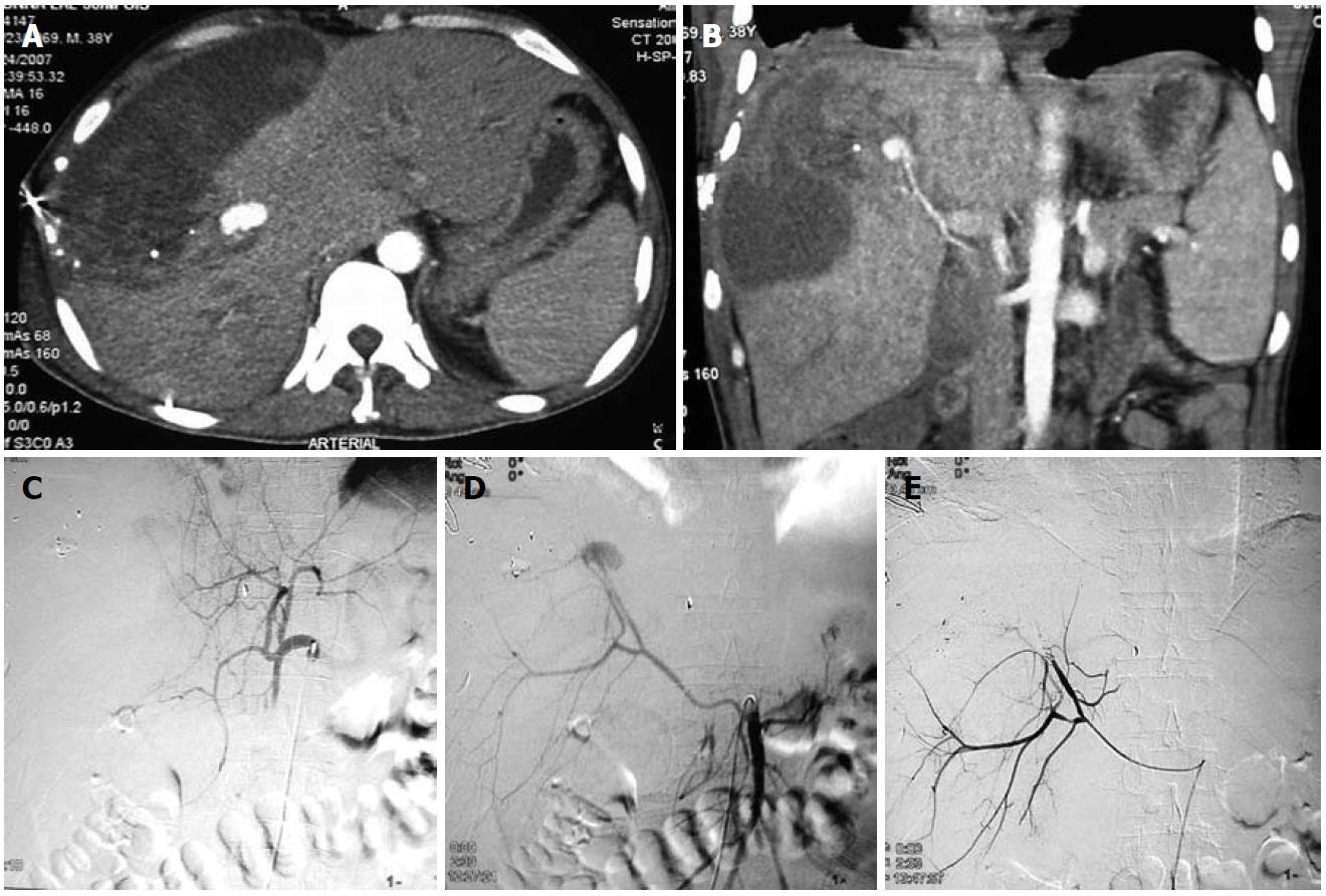
Figure 2 Traumatic pseudoaneurysm of a replaced right hepatic artery.
A 38-year-old male presented with hypotension and falling hematocrit after a road traffic accident. A, B: Axial contrast-enhanced computed tomography of the abdomen (A) and coronal MIP image (B) revealed a large pseudoaneurysm (arrows) in the right lobe of the liver and a large hematoma in the liver parenchyma extending to the subcapsular location; C: DSA image after selective catheterization of the celiac trunk failed to reveal any aneurysm; D: Selective superior mesenteric artery (SMA) catheterization revealed a replaced right hepatic artery arising from the SMA and a pseudoaneurysm arising from it; E: Treated with coil embolization.
- Citation: Jana M, Gamanagatti S, Mukund A, Paul S, Gupta P, Garg P, Chattopadhyay TK, Sahni P. Endovascular management in abdominal visceral arterial aneurysms: A pictorial essay. World J Radiol 2011; 3(7): 182-187
- URL: https://www.wjgnet.com/1949-8470/full/v3/i7/182.htm
- DOI: https://dx.doi.org/10.4329/wjr.v3.i7.182


