Copyright
©2011 Baishideng Publishing Group Co.
World J Radiol. Jul 28, 2011; 3(7): 169-177
Published online Jul 28, 2011. doi: 10.4329/wjr.v3.i7.169
Published online Jul 28, 2011. doi: 10.4329/wjr.v3.i7.169
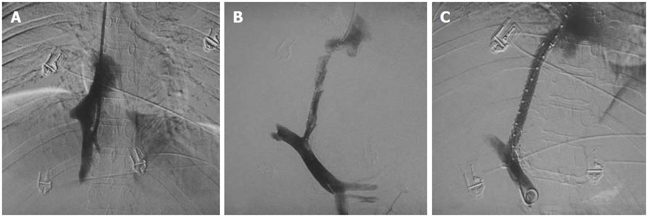
Figure 10 DSA images of chronic Budd-Chiari syndrome with complete occlusion of all hepatic veins, cavogram.
A: Small stump of right hepatic vein; B, C: Creation of porto-caval shunt and placement of transjugular intrahepatic portosystemic shunt stent.
- Citation: Mukund A, Gamanagatti S. Imaging and interventions in Budd-Chiari syndrome. World J Radiol 2011; 3(7): 169-177
- URL: https://www.wjgnet.com/1949-8470/full/v3/i7/169.htm
- DOI: https://dx.doi.org/10.4329/wjr.v3.i7.169


