Copyright
©2011 Baishideng Publishing Group Co.
World J Radiol. Jul 28, 2011; 3(7): 169-177
Published online Jul 28, 2011. doi: 10.4329/wjr.v3.i7.169
Published online Jul 28, 2011. doi: 10.4329/wjr.v3.i7.169
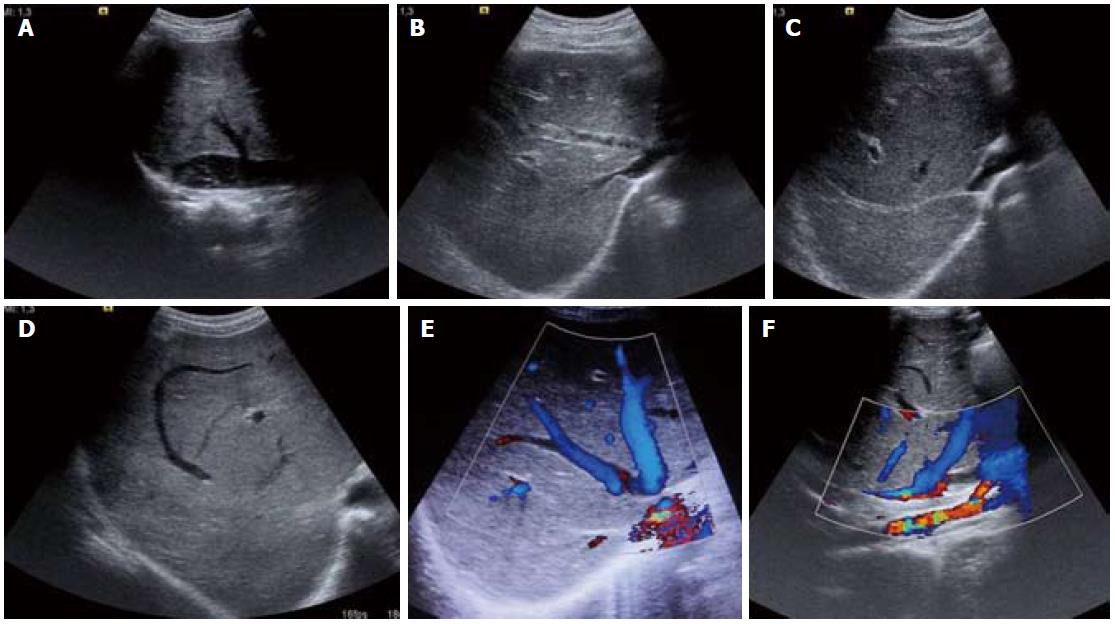
Figure 2 Ultrasound images (A-D) and color Doppler images (E, F).
A: Inferior vena cava web with intra-luminal floating thrombus; B: Partial thrombus within the middle hepatic vein and osteal narrowing of the right hepatic vein; C: Fibrosed right hepatic vein; D: Comma-shaped intrahepatic collaterals; E, F: Web at hepatic vein ostium and intrahepatic collaterals.
- Citation: Mukund A, Gamanagatti S. Imaging and interventions in Budd-Chiari syndrome. World J Radiol 2011; 3(7): 169-177
- URL: https://www.wjgnet.com/1949-8470/full/v3/i7/169.htm
- DOI: https://dx.doi.org/10.4329/wjr.v3.i7.169


