Copyright
©2011 Baishideng Publishing Group Co.
World J Radiol. May 28, 2011; 3(5): 125-134
Published online May 28, 2011. doi: 10.4329/wjr.v3.i5.125
Published online May 28, 2011. doi: 10.4329/wjr.v3.i5.125
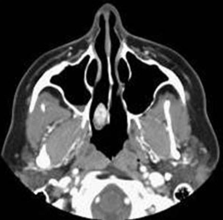
Figure 9 Hemangioendothelioma.
Axial computed tomography shows a non-specific sclerotic enhanced lesion related to the posterior part of the nasal septum on the right side (Courtesy of Castillo M).
- Citation: Razek AAKA. Imaging appearance of bone tumors of the maxillofacial region. World J Radiol 2011; 3(5): 125-134
- URL: https://www.wjgnet.com/1949-8470/full/v3/i5/125.htm
- DOI: https://dx.doi.org/10.4329/wjr.v3.i5.125


