Copyright
©2011 Baishideng Publishing Group Co.
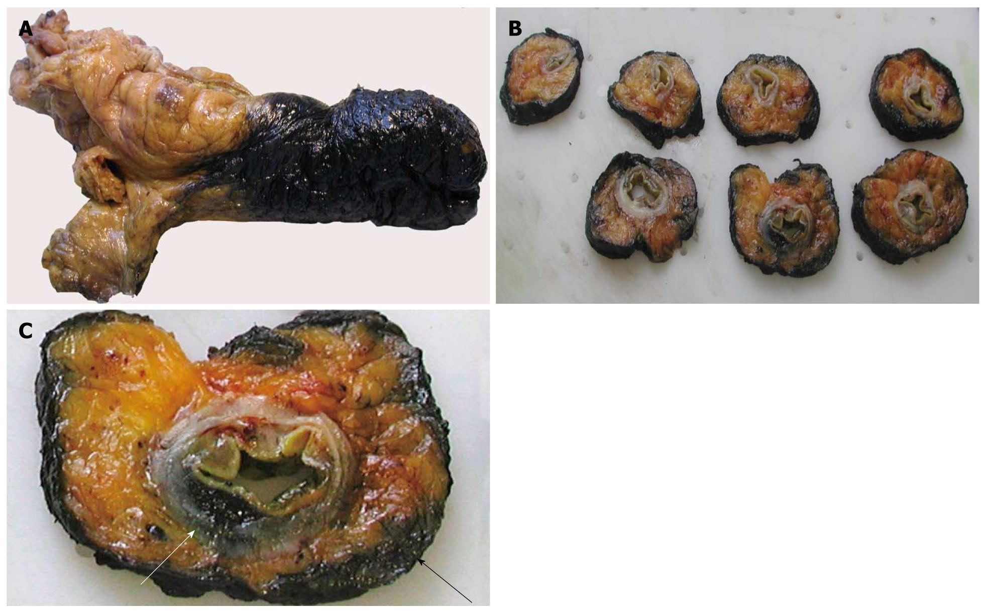
Figure 3 Pathological evaluation of the circumferential resection margin (CRM).
A: The excised rectum is dipped in ink; B: Serial sections including the tumor are taken through the entire rectum; C: One of the sections shows the tumor’s leading edge (white arrow) and its relation with the CRM (black arrow).
- Citation: Bellows CF, Jaffe B, Bacigalupo L, Pucciarelli S, Gagliardi G. Clinical significance of magnetic resonance imaging findings in rectal cancer. World J Radiol 2011; 3(4): 92-104
- URL: https://www.wjgnet.com/1949-8470/full/v3/i4/92.htm
- DOI: https://dx.doi.org/10.4329/wjr.v3.i4.92


