Copyright
©2011 Baishideng Publishing Group Co.
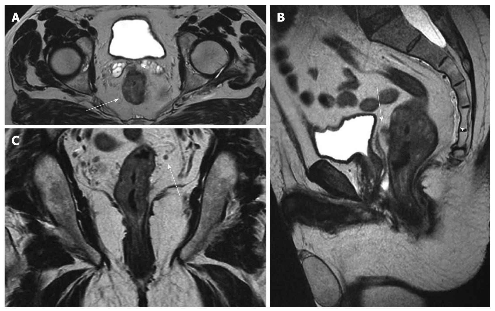
Figure 1 Magnetic resonance imaging staging of rectal cancer before chemoradiation: T3, N+.
Pathology result: T3, N1. A: Axial T2w image shows the circumferential tumor (arrow) and the extramural spread anteriorly (arrowhead) close to the seminal vesicles; B: In the sagittal T2w image, the anterior extramural spread (arrow) can be also recognized close to the mesorectal fascia (thin vertical hypointense line posterior to the bladder). The presacral fascia can also be appreciated (arrowhead) continuing inferiorly as the rectosacral fascia; C: In the coronal T2w image, a small mesorectal lymph node (arrow) is seen.
- Citation: Bellows CF, Jaffe B, Bacigalupo L, Pucciarelli S, Gagliardi G. Clinical significance of magnetic resonance imaging findings in rectal cancer. World J Radiol 2011; 3(4): 92-104
- URL: https://www.wjgnet.com/1949-8470/full/v3/i4/92.htm
- DOI: https://dx.doi.org/10.4329/wjr.v3.i4.92


